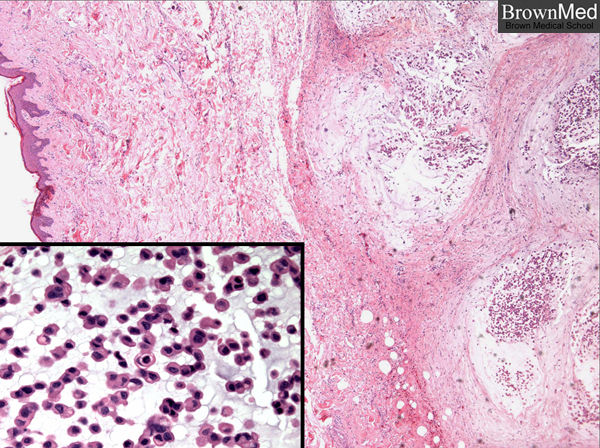
Chordoma
This midline tumor arises from notochordal elements. These photos are from a recurrent chordoma of the spine and left flank in a 68
year old man. The specimen contained gelatinous yellow and pink-tan fragments of tissue and a resected scar with tumor infiltrating
the subcutaneous tissue. Microscopically, lobules of tumor are separated by fibrous septa. Cords of tumor cells are seen in a myxoid background. Vacuoles can be seen and when prominent, the cells are referred to as physaliferous (Gk. physali bladder, L. ferre to bear).
With thanks to Dr. Teresita Redondo, St. Barnabas Medical Center, Livingston, NJ.