Sarcoidosis
A 46 year old black man with a history of lupus erythematosus, positivity for hepatitis C, and renal failure with several
years on dialysis entered with fever and dyspnea. He was found to have bilateral pulmonary infiltrates, hilar adenopathy,
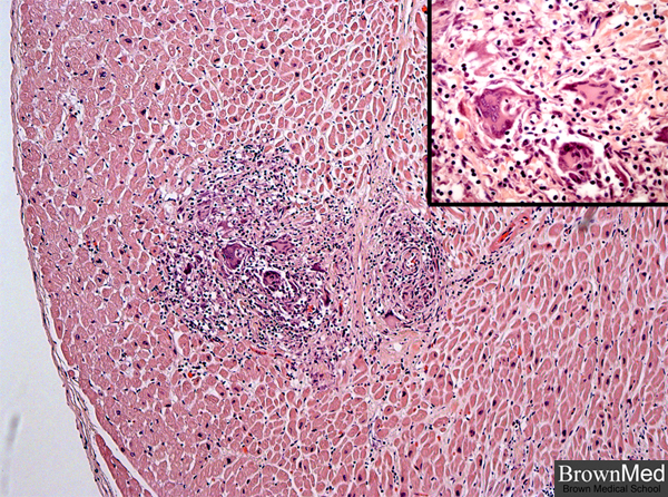
and cardiomegaly. This photo of the myocardium reveals two non-caseating granulomas with giant cells, consistent with
sarcoidosis. The inset offers a higher magnification view of giant cells. Pre- and postmortem studies for tubercle bacilli and
fungi were negative. For more photos from this interesting case see pulmonary and lymph-related sections on this website.
Contributed by Dr. Robert Van Wesep
1 minute clinical correlation