Rhabdomyoma
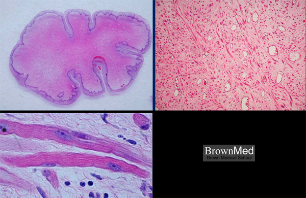
A 46 year old woman was found to have an incidental 2-3 cm vaginal polyp at the time of hysterectomy for uterine
fibroids. The upper left photo shows a polypoid lesion covered by normal vaginal epithelium. In the upper right photo the
lesion consists of loose connective tissue, thin-walled blood vessels, and a vaguely fascicular proliferation of strap cells; the
latter differentiating to skeletal muscle. In the lower photo, taken at high magnification and with the condenser lowered,
cross-striations are readily seen. The histology is consistent with a benign lesion; no atypia or mitoses were present.
Contributed by Dr. Dan Koelliker
1 minute clinical correlation