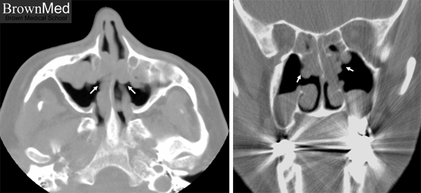
Sinonasal inflammatory polyp
Axial (left) and coronal (right) view of CT imaging of the face demonstrating multiple nasal polyps (arrows)
that appear to have originated from both the lateral wall and the ethmoid process. The patient who has had
prior nasal polypectomy presented with chronic sinusitis, nasal obstruction and mild headache.
1 minute clinical correlation