 Laboratory Primate Newsletter
Laboratory Primate Newsletter
Laboratory Primate Newsletter VOLUME 26 NUMBER 3 JULY 1987
Articles and Notes
Improved Installation Method for Branches as Cage Enrichment, by V. Reinhardt ...... 1
B-Virus Infection in Humans ...... 2
Captive Breeding of Alouatta belzebul and Chiropotes satanus utahicki, by W. R. Kingston ...... 8
News, Information, and Announcements
The Use of Nonhuman Primates in Laboratories in Great Britain ...... 4
Encephalitozoon cuniculi Infection Detected in Squirrel Monkeys ...... 5
ABS/ASAB Guidelines for the Use of Animals in Research ...... 6
Meeting Announcement: IPS ...... 7
Erratum ...... 7
Letters: Primate Herpes Virus ...... 8
Tarsier Research Opportunity ...... 8
Pathology of Laboratory Animals ...... 18
Departments
Recent Books and Articles ...... 9
* * *
Viktor Reinhardt
University of Wisconsin
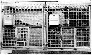
We have recently commented on the usefulness of deciduous tree branches for enriching the environment of singly caged rhesus monkeys (Reinhardt, et al., 1987). During that 2-month pilot study, we installed branches without attachment in the cage. We did not have to remove any branches for hygienic reasons, but it became clear that special care is needed during routine cage cleaning to prevent food and fecal material from accumulating in the corner where the lower end of the branch touches the floor of the cage.
To save extra cleaning time, while simultaneously ensuring optimal hygienic conditions in the cage, we have devised a simple technique of branch installation. As shown in Figure 1, the branch is now suspended. Its lower end is attached with a chain at the front of the cage, while its upper end rests at the junction of the back and side wall without touching the top grating of the cage. This installation makes it impossible for food and fecal material to accumulate in the cage due to the branch. Special cleaning is therefore no longer necessary.
We attach the branch in such a way that the animal can both perch on it and freely move below it. Usable cage space is thus maximized for the animal.
We have tested the new installation method in 272 cases over a period of 30 days and are confident that we have found an optimal way to eliminate the need of special cage cleaning due to the branch.
Figure 1: Suspended installation of the branch increases the usable cage space for the monkey and makes it impossible for food and fecal material to accumulate at the lower end of the branch.
Reference
Reinhardt, V., Houser, W. D., Cowley, E., & Champoux, M. (1987). Preliminary comments on environmental enrichment with branches for individually caged rhesus monkeys. Laboratory Primate Newsletter, 26(1), 1-3.
-------------------------------------------------------------------
Author's address: Wisconsin Regional Primate Research Center, 1223 Capitol Court, Madison, WI 53715-1299.
This projected was supported by NIH grant RR00167 to the Wisconsin Regional Primate Research Center.
-------------------------------------------------------------------
* * *
Between March 28 and April 7, 1987, four persons were admitted to hospitals in Pensacola and Gulf Breeze, Florida, with illnesses that were later confirmed to be caused by infection with B-virus (cercopithecid herpesvirus 1, Herpesvirus simiae [Matthews, 1982]). Three were monkey handlers with the Naval Aerospace Medical Research Laboratory (NAMRL) at the Pensacola Naval Air Station; the fourth was the wife of one of the three handlers.
Patient 1: About March 4, a 31-year-old male who had been employed as an animal caretaker for 8 years was bitten on the left thumb by a 3-year-old rhesus monkey that was suffering from severe bilateral conjunctivitis and diarrhea. The employee had occasionally handled smaller monkeys without protective leather gloves, and it is not certain whether he was wearing gloves when he was bitten. Five days later, he developed numbness in his left arm. Eighteen days after being bitten, he developed lethargy, fever, chills, dizziness, and myalgia. At no time did he have skin lesions suggestive of herpesvirus infection. Over the next 4 days, he developed numbness and paresthesia in the left side of his body, diplopia, and leg weakness. On March 28, he was admitted to the hospital. Two days later, he was placed on intravenous acyclovir. Subsequently, B-virus antibodies were detected in his serum by enzyme immunoassay (titer = 32). Spinal fluid that was collected before the initiation of acyclovir therapy was positive for B-virus. The patient continued to deteriorate and was put on a respirator. His therapy was changed to 9-(1.3-dihydroxy-2-propoxymethyl)-guanine (DHPG) on a compassionate Investigational New Drug protocol granted by the Food and Drug Administration. He is currently semi-comatose.
Patient 2: On or about March 10, a 37-year-old male who had been employed as a biological technician for 13 years suffered a penetrating wound which may have been a monkey bite or scratch on the left forearm. Patient 2 had had frequent contact with the monkey that injured Patient 1, and his wound may have been inflicted by this animal. Patient 2 had also handled smaller animals without leather gloves, but it is uncertain whether he was wearing them at the time he was exposed. Five days after his injury, he developed herpetiform vesicles at the site of the wound. On March 26, after the lesions had become crusted, he was seen by a dermatologist who detected giant cells in scrapings from the lesions (Tzanck preparation) but no distinct viral inclusions. A presumptive diagnosis of herpes zoster versus herpes simplex was made. Topical acyclovir was prescribed, but the patient treated himself only with topical hydrocortisone cream. Over the next several days, he developed numbness in his left arm, chest pain, dyspnea, fever, confusion, lethargy, diplopia, and dysphagia. He made several visits to emergency rooms before being hospitalized on March 28. Later that day, he suffered a respiratory arrest and was placed on mechanical ventilation. A lumbar puncture was consistent with aseptic meningitis. He was placed on intravenous acyclovir. A skin biopsy specimen obtained the day after admission was positive for B-virus. Treatment was subsequently changed to intravenous DHPG. However, the patient's condition deteriorated, and he died on April 28.
Patient 3: On March 11, a 53-year-old male laboratory supervisor who had been employed at NAMRL for 12 years handled a clinically healthy monkey. He wore leather gloves to catch the animal but wore only surgical gloves while holding it afterward. He reported no bites, scratches, or contact with monkey body fluids. On March 27, he noted pruritic vesicles on the third finger of his right hand. Three days later the lesions were dry and crusted. A physician at the laboratory referred him to a dermatologist who performed a biopsy and later placed him on oral acyclovir. The tissue obtained during the biopsy was positive for a herpesvirus, and, on April 6, the patient was hospitalized. Intravenous acyclovir was begun on April 10, and the tissue was confirmed positive for B-virus on April 13. The lesions continued to heal, and the disease did not progress further. On April 21, the patient was discharged from the hospital and instructed to continue treatment with oral acyclovir. However, he greatly reduced his dosage a few days later. Routine follow-up cultures of conjunctiva and buccal mucosa obtained on April 28 were positive for B-virus the following day. He was readmitted to the hospital and again placed on intravenous acyclovir. He has remained asymptomatic. All other follow-up cultures except a rectal culture obtained May 8 have been negative.
Patient 4: The 29-year-old wife of Patient 2 applied hydrocortisone cream to her husband's skin lesions beginning about March 18. During this time, she also applied this cream to an area of contact dermatitis under a ring on her finger. The dermatitis was highly pruritic, and she scratched it to the point of bleeding. On April 1, she was seen by a dermatologist who performed a culture of samples taken from the lesion and prescribed oral acyclovir. On April 7, the culture was reported positive for B-virus, and the patient was hospitalized and placed on intravenous acyclovir. Her dermatitis cleared, and the disease did not progress further. Cultures of oral and conjunctival specimens were performed every 3 to 4 days. The conjunctival cultures became positive for B-virus beginning with the specimen of April 10 and remained positive through April 28. She had no clinical evidence of conjunctivitis, and subsequent cultures have been negative.
Forty-nine persons who had direct (skin-to-skin or body-fluid-to-skin) contact with the patients before diagnosis are under clinical and laboratory surveillance for B-virus infection. No cases of infection or illness suggestive of B-virus have been detected among this group. The ill monkey that bit Patient 1 and that may have bitten Patient 2 and the clinically healthy monkey that was handled by Patient 3 have positive saliva cultures for B-virus.
Note: B-virus, a close relative of the herpes simplex viruses of man, is enzootic in macaques and possibly other Old World monkeys. It is most frequently associated with rhesus monkeys (Macaca mulatta). Like herpes simplex virus infections in man, B-virus infection in monkeys is characterized by intermittent reactivation and shedding, particularly during periods of stress and/or immunosuppression. Fortunately, symptomatic infection in monkey handlers and in persons handling monkey tissue appears to be rare--since the discovery of the virus in the 1930's, only 23 cases of symptomatic human infection have been described in the literature (Palmer, 1987). However, the consequences of symptomatic infection are severe--of the 23 patients, 18 have died from encephalitis. The frequency of asymptomatic human infection is unknown.
In at least one instance, Patient 1 and Patient 2 had handled an ill monkey that had not been anesthetized. It appears that at least one of them had not worn the recommended protective clothing. One was bitten, and the other was either bitten, scratched, or infected through contamination of a preexisting wound. It is, therefore, likely that the use of appropriate protective clothing could have prevented illness in at least one of the men. Patient 3, however, was appropriately protected when he handled the second culture-positive monkey, and he was not aware of any skin contact with the monkey or its body fluids. However, he may have had unrecognized contact with contaminated material.
Patient 4 has the first documented case of human-to-human transmission of B-virus. Infectious fluid from her husband's skin lesions was apparently inoculated directly into macerated skin, similar to the inoculation produced by a monkey bite. Since her infection does not appear to have spread systemically, she may have spread the infection to her eyes when she inserted her contact lenses. Transmission of the virus by less direct contact, such as inoculation of infectious fluid on intact skin or transmission by fomites, although theoretically possible, has not been documented. The lack of detectable infection thus far in persons with such exposures to any of the four patients suggests that transmission from casual contact is unlikely. This information will be important as public health recommendations are developed for releasing Patient 3 and Patient 4 back into the community.
This outbreak serves as a reminder of the inherent risk in working with macaques and possibly with other Old World monkeys. These monkeys should not be used for research purposes unless the handlers can adhere strictly to published guidelines. These guidelines state that persons working with macaques should wear gloves and laboratory coats to avoid bites and scratches (CDC, 1984). To further reduce risk, monkeys, especially large ones, should be anesthetized before handling, when it is feasible, or should be housed in squeeze cages.
The most important control measure is the careful education of animal caretakers and laboratory personnel who handle monkey tissues. The following points should be emphasized: 1) the nature and risk of B-virus infection, 2) the need to rapidly and thoroughly cleanse any penetrating wounds, 3) the need to seek medical attention immediately if suspicious lesions or other symptoms such as intense pruritus or numbness occur, and 4) the need for any physician suspecting B-virus infection to consult public health authorities and to institute appropriate diagnostic and therapeutic measures. So far, acyclovir therapy appears to have prevented the progression of disease in Patient 3 and Patient 4. The apparent responsiveness of these infections to treatment underscores the importance of early recognition and treatment of B-virus infection in sympotomatic persons.--[From Morbidity and Mortality Weekly Report, 36, 1987, 289-296.]
References
Matthews, R. E. F. (1982). Clssification and nomenclature of viruses. Fourth Report of the International Committee on Taxonomy of Viruses. New York: S. Karger. P.49.
Palmer, A. E. (1987). B-virus, Herpesvirus simiae: historical perspective. Journal of Medical Primatology, 16, 99-130.
CDC, National Institutes of Health (1984). Biosafety in microbiological and biomedical laboratories. Bethesda, MD: US Dept of Health and Human Services, Public Health Service. DHHS publication no. (CDC)84-8395.
* * *
One justification for the use of primates in biomedical research is their close evolutionary relationship to humans. Those parts of the brain associated with general awareness and appreciation of distress and suffering are more highly developed in primates than in lower mammals. In other words, according to the Fund for the Replacement of Animals in Medical Experimentation (FRAME) and the Committee for the Reform of Animal Experimentation (CRAE), the stronger the physiological criteria for using nonhuman primates as models for human beings in laboratory experiments, the stronger the ethical or humanitarian reasons for not doing so. FRAME and CRAE have drawn up a report (The Use of Non-human Primates as Laboratory Animals in Great Britain. Available from FRAME, Eastgate House, 34 Stoney St., Nottingham NG1 1NB [0602 584740]) which points out the error of assuming that primates are the most appropriate animal model--in many instances the responses of rodents have been closer to those of human beings. FRAME and CRAE are concerned about the welfare of all animals used in laboratory procedures, while recognizing that biomedical research may not only reduce human suffering but also reduce animal suffering. The report draws attention to the high levels of stress and abnormal behaviour that can occur in laboratories: when family units are divided (particularly mother and baby); when primates are kept in isolation; when they are maintained in small cages; when they can anticipate repeated painful or unpleasant procedures; and when there are no facilities for privacy or recreation. Many primates in laboratories are captured in the wild and may be subject to poor conditions during long journeys to other countries. Although breeding can take place successfully in laboratories, the authorities of the report are concerned that primates may then be used for general purposes simply because they are available. FRAME and CRAE urge special classification of primates in the annual returns to the Home Office; that primates should be subjected to mild procedures only (excluding routine toxicity tests) in research; and that a code of good practice should be drawn up to reduce the number of primates used in laboratories and to minimize their pain and suffering.--From The Lancet, January 31, 1987.
* * *
Several serological tests have been developed and refined to detect a parasitic organism that can seriously confound interpretation of experimental data obtained from laboratory animals.
"Encephalitozoon cuniculi is a severe contaminant of certain laboratory animals, and its impact is seriously underestimated by many investigators," Dr. John Shadduck of the University of Illinois notes. The clinically inapparent, persistent infections by the parasite have been found in up to 75 percent of laboratory rabbits and up to 50 percent of laboratory rats and mice in some closed colonies. Scientists recently found the naturally occurring infection in a colony of squirrel monkeys.
E. cuniculi are shed in the urine and invade animals primarily through nasal-oral routes, Dr. Shadduck says. Placental transmission of the parasite also is possible.
At the Delta Regional Primate Research Center (PRC) in Covington, LA, the threat of E. cuniculi infection is taken very seriously, according to Dr. Gary B. Baskin, head of the pathology department at the Center. "E. cuniculi may have led to several spontaneous abortions and neonatal deaths in our squirrel monkey population. I've seen sporadic cases over the last few years, and we see a flurry of severe cases every spring when the infants are born."
During the recent 20-month period, 17 percent of the squirrel monkeys examined at necropsy were harboring Encephalitozoon cuniculi. Infected squirrel monkeys characteristically showed lesions in the brain, kidney, lung, adrenal, liver, and placenta. Inflammation of blood vessels and aorta also was seen, according to Dr. Baskin.
Since the initial 20-month study, the researchers have screened approximately 240 animals with serological tests. The investigators report that about 75 percent of the monkeys have had, at one time or another, a positive antibody titer to the protozoan.
Dr. Baskin speculates that the monkey colony was likely infected by wild rodents that entered the PRC grounds. Interestingly, although several different species of monkeys at the PRC have outdoor housing facilities, only squirrel monkeys appear to harbor E. cuniculi.
"We have several thousand monkeys in outdoor colonies, and a few hundred of these are squirrel monkeys," Dr. Baskin says. "All these animals have an abundant opportunity to come into contact with infected rodents. Why we see encephalitozoonosis so often in the squirrel monkeys and never in rhesus monkeys, for example, we just don't know."
The difference in susceptibility to E. cuniculi infection is maintained among and within species of animals, as seen by the various responses in different monkey colonies at the PRC, Dr. Shadduck points out. Other animals, such as dogs and cats, either succumb to the infection and die, or clear the protozoan from the body. Rabbits and mice, on the other hand, maintain a persistent but inapparent infection. "The presence of these differences poses one of the most interesting questions in E. cuniculi studies. It is also very intriguing that, as far as we can tell, the parasite is identical in all species. We think that differences in responses among animals are not due to the parasite, but rather to the host."
The mechanism by which all infected animals--rats, mice, rabbits, and monkeys--kill the parasite seems to be the same. The protozoan stimulates T cells, which release some intermediate factor, most likely a lymphokine. Lymphokines, in turn, activate macrophages, which kill the parasites. Precisely what macrophages do to kill E. cuniculi is not known.
Dr. Baskin recommends screening squirrel monkeys because the animals appear to be very susceptible to E. cuniculi. "It's difficult to impress upon researchers how often a study is confounded or confused by this parasite." --[Excerpted from the Research Resources Reporter, 11[5], 1987, 9-11]
* * *
The Animal Behavior Society and the Association for the Study of Animal Behaviour have formed Animal Care and Ethical Committees respectively. These committees jointly produced guidelines for the use of those who are planning and conducting studies of animal behavior. These guidelines will be used by the editors of Animal Behaviour. Submitted papers that appear to violate the spirit of the guidelines will be referred to one of the committees, and the evaluation of the committee will be used by the editor in deciding whether to accept the manuscript. An outline of the guidelines follows:
1. Investigators must abide by the spirit as well as the letter of relevant legislation.
2. The species chosen for study should be well-suited to answer the questions posed. When research involves the use of procedures that are likely to cause unavoidable pain or discomfort to the animal, and when alternate species can be used, the researcher should employ the species which in the opinion of the researcher and other qualified colleagues is least likely to suffer.
3. In laboratory studies or field studies involving manipulations potentially detrimental to the animal or the population, the researcher should use the smallest number of animals necessary and sufficient to accomplish the research goals.
4. If procedures used in research involve pain or discomfort, the investigator must consider whether the knowledge that may be gained justifies the stress and pain inflicted on the animals. In general, researchers are urged to consider the use of alternative procedures before employing techniques that are likely to cause physical or psychological discomfort to the animal. If aversive stimulation or deprivation is used, the investigator should ascertain that there is no alternative way of motivating the animal, and that the levels of deprivation or aversive stimulation used are no higher than necessary to achieve the goals of the experiment. Experimental designs which require keeping animals in over-crowded conditions, or which involve social deprivation or isolation, may be extremely stressful to the animals involved. Where feasible, studies inducing a deleterious condition in animals should also address the possible treatment, prevention, or alleviation of the condition.
5. Members of endangered or locally rare species should not be collected or manipulated in the wild except as part of a serious attempt at conservation.
6. Animals should be obtained only from reliable sources. So far as is possible, the investigator should ensure that those responsible for handling the animals en route to the research facilities provide adequate food, water, ventilation and space, and do not impose undue stress. If animals are captured or killed in the wild this should be done in as painless and humane a manner as possible.
7. The experimenter's responsibilities extend also to the conditions under which the animals are kept when not in use. Caging conditions and husbandry practices must meet, at the very least, minimal recommended requirements.
8. Whenever practical or feasible, researchers should attempt to distribute their animals to colleagues for further study, but care should be taken that the same animals are not used repeatedly in experiments which involve invasive surgical procedures or other treatments which are likely to be stressful or painful. If animals must be destroyed subsequent to a study, this should be done in as humane and painless a way as possible, and death of the animals should be confirmed before their bodies are discarded.
These guidelines should not be considered an imposition upon the scientific freedom of individual researchers, but rather as helping to provide an ethical framework to which each investigator may respond in making decisions related to animal welfare.
* * *
The Organizing Committee of the XIIth Congress of the International Primatological Society is pleased to invite members of the IPS and all persons interested in the various fields of Primatology, to participate in the Congress, which will be held in the Brasília Convention Center, Brasília, Brazil, on July 24-29, 1988.
Primatological activities in Brazil have been intense in the last years, due in great part to the foundation of the Brazilian Society of Primatology, in 1979. Primatologists and other interested persons from all over the world will have the opportunity of exchanging experiences with their Brazilian colleagues. There will be chances to visit active groups in different parts of the country.
The official language of the Congress will be English, with simultaneous translation to Portuguese and Spanish in the symposia and conferences. The scientific activities will include an opening session, lectures, symposia, conferences, workshops, paper sessions, poster presentations, films, and video tapes. Emphasis will be given to poster presentations because they facilitate discussion, perusal of data, and one-to-one exchange of ideas. If you are interested in organizing a symposium and still have not informed the Committee, please send details immediately.
Social functions and post-Congress field excursions are planned. Because of Brasília's central location it is easy to travel to other cities in Brazil, such as Rio de Janeiro, São Paulo, and Salvador, and to other Latin American countries.
Excursions to Pantanal and Amazonia will be organized by André Safari Tours. Since the number of participants in each excursion must be limited it is advisable to contact André Safari as soon as possible (Address: SHI Sul, QI 7, Bl. B, Room 201, P.O. Box 7020, CEP 71619, Brasília DF, Brasil. Telephone: 061-248-3953).
VARIG Airline is the official carrier for the Congress. There will be special treatment and travel arrangements for the delegations. Further information is available at local VARIG offices throughout the world. Due to the great number of tourists to Brazil, we suggest contacting the nearest VARIG agent as soon as possible for flight reservations, Brazil Air Pass, and flight/hotel packages.
For extensive travel in Brazil before or after the Congress it is convenient to buy a low priced Brazil Air Pass. It may be purchased outside Brazil by foreigners and Brazilians who are residents of a foreign country. VARIG Airline offices can give more information. Local transportation in Brasília will be provided, including between hotels and the Convention Center.
The deadline for submission of abstracts and hotel reservations will be December 31, 1987. For detailed information, write to: The Organizing Committee, XIIth Congress of the International Primatological Society, c/o Prof. Milton Thiago de Mello, Departamento de Biologia Celular, Instituto de Ciências Biológicas, Universidade de Brasília, 70910, Brasília, DF, Brasil. [Telephone (061) 274-0022, extension 2176. Telex (061) 2730 UNBS BR]
* * *
In the article "The African green monkey (Cercopithecus aethiop sabaeus) as a carrier of diseases on Barbados," by J. Baulu, C. O. R. Everard, and J. D. Everard (Laboratory Primate Newsletter, 26(2), 1987, 2-4), the first sentence of the last paragraph on page 3 should read as follows: During the course of handling several thousand wild-caught monkeys, we observed at least 100 adults with moderate to severe oral problems.
* * *
Just a note to say that I have retired and been replaced by Professor C. A. Hart. He and I will continue our research work on New World primate herpes viruses. In particular we are using Herpesvirus saimiri (otherwise H. tamarinus, or platyrhine herpes, or marmoset herpes), now properly called HVS1 (for Herpesvirus saimiri 1) as a model for studying human herpes simplex encephalitis (called HSV1, thus very confusing).
We have several strains of the virus, some of which are virtually nonvirulent in the rabbit but which always produce latent infection in dorsal rod ganglion cells; we have one highly virulent strain with only minor but no doubt important differences in R. E. pattern of enzyme digests of DNA. We will be looking closely at possible gene products, etc.--Kevin McCarthy, Dept. of Medical Microbiology, Duncan Building, Royal Liverpool Hospital, P.O. Box 147, Liverpool L69 3BX, Great Britain.
* * *
The Duke University Primate Center holds colonies of two species of living tarsiers. The Duke Primate Center is a national research facility available to all interested researchers for short- or long-term studies on the behavior, anatomy and biochemistry of prosimians. Inquiries about research on Duke's tarsiers should be directed to Elwyn L. Simons, Director, Duke University Primate Center, 3705 Erwin Road, Durham, NC 27705 [Telephone: (919) 489-3364].
* * *
W. R. Kingston
Centro Nacional de Primatas, Belém, Brazil
In my note on the behavior of the above two species (Kingston 1985), I mentioned that breeding groups had been set up in the Brazilian National Primate Centre in Belém, Brazil. I have recently been advised by the director, Dr. Jose Muniz, that a number of Alouatta belzebul and at least one Chiropotes satanus utahicki have been born and are being maintained. We believe that these are the first captive breedings of both species. A number of young Howlers were born to females which were pregnant when rescued from the Tucurui dam flooding but, not surprisingly, these did not survive. The births which are now occurring in both species are from animals which have been in captivity well over two years.
A point of great satisfaction to me is that the original female Alouatta belzebul, "Preta," received as a very young orphan in March 1981, and which had been kept singly and become extremely tame, has not only produced one thriving youngster but is again pregnant at the time of writing. She was extremely hostile to a carefully selected male from the Tucurui animals when they were introduced early in 1985, and when I left Brazil in May of that year breeding seemed extremely doubtful. It appears, however, that the tameness and possible imprinting on humans have not prevented normal social behavior after all.
Reference
Kingston, W. R. (1985). Note on the behavior of the "difficult" neo-tropical primate genera in captivity. Laboratory Primate Newsletter, 24(1), 10-11.
-------------------------------------------------------------------
Author's address: The Old Smithy, Bishops Frome, Worcester WR6 5BA, England.
-------------------------------------------------------------------
* * *
Books
*West Indian Green Monkeys: Problems in Historical Biogeography. Contributions to Primatology, Vol. 24. Basel: Karger, 1987. 80 pp. [Price: $22.251.
. . .
The author synthesizes 350 years of documentary evidence in order to better understand important facets of the history of West Indian green monkeys. In so doing, he seeks to demonstrate the importance of using historical documents to reconstruct primate history and so obtain knowledge on the evolution, geographical distribution, genetic composition and population properties of an introduced primate species. Two major issues are explored: the migration of African green monkeys to Barbados, St. Kitts, and Nevis, and changes in the Barbadian monkey population during the last 350 years. The book is not intended as a definitive history, but rather as a stimulus and guide to future research. The problems remain unsolved, but several competing and testable hypotheses are proposed as solutions for each of them.
* Primates: The Road to Self-Sustaining Populations. Kurt Benirschke (Ed.). New York: Springer-Verlag, 1986. 1044 pp. [Price: $79.00].
. . .
The proceedings of a conference held in San Diego, CA, June 24-28, 1985, sponsored by The Morris Animal Foundation, Englewood, CO, and The Zoological Society of San Diego, San Diego, CA. Contents: Foreword by J. Diamond; Primate ethics, by R. V. Short. The role of captive populations in global conservation, by D. Western. African primate conservation: General needs and specific priorities, by J. F. Oates. Gorilla conservation: Anatomy of a campaign, by A. H. Harcourt. Captive chimpanzee populations--past, present, and future, by U. S. Seal & N. R. Flesness. Responses of rainforest primates to selective logging in Kibale Forest, Uganda: A summary report, by J. P. Skorupa. Lemur survival, by A. Jolly. The conservation status of nonhuman primates in Indonesia, by K. MacKinnon. Southeast Asian primates, by D. J. Chivers. Conservation of orangutans: A status report, 1985, by H. D. Rijksen. The natural breeding strategy of gibbons (Hylobates lar); Are we managing the captive population by design or default?, by D. E. Moore. The primates of India: Status, trends, and conservation, by C. H. Southwick & D. G. Lindburg. Increased home range for a self-sustaining free-ranging rhesus population at Tughlaqabad, India, by I. Malik. Balancing the wild/captive equation--the case of the Barbary macaque (Macaca sylvanus L.), by J. E. Fa. Primate status and conservation in China, by S. Wang and G. Quan. Primate conservation priorities in the neotropical region, by R. A. Mittermeier. Ecological background and conservation priorities for woolly spider monkeys (Brachyteles arachnoides) , by K. Milton. Successes and failures of captive breeding, by M. L. Jones. History of Geoffroy's tamarins, Saguinus geoffroyi, at Lincoln Park Zoological Gardens; 1974-1985, by D. A. Meritt, Jr. The management of prosimians in captivity for conservation and for research, by J. I. Pollock. Corral breeding of nonhuman primates, by W. J. Goodwin. Environments for captive propagation of primates: Interaction of social and physical factors, by J. Erwin. Behavior requirements for self-sustaining primate populations--some theoretical considerations and a closer look at social behavior, by J. A. R. A. M. van Hooff. Early socialization, by W. A. Mason. Behavioral aspects of successful
reproduction in primates, by S. J. Suomi. Conflict resolution in monkeys and apes, by F. B. M. de Waal. Resocialization of asocial chimpanzees, by J. Fritz. Primate mating systems and their consequences for captive management, by R. L. Tilson. The interbirth interval in primates: Effects of pregnancy and nursing, by R. F. Williams. Embryonic loss in primates in relation to in vitro fertilization and embryo transfer, by G. D. Hodgen. Artificial acceleration of reproduction, by J. P. Hearn. Collection, assessment, and storage of sperm, by W. V. Holt. Artificial insemination of nonhuman primates, by K. G. Gould & D. E. Martin. Primate models for fertilization and early embryogenesis, by W. R. Dukelow & P. N. Vengesa. Housing and furniture, by W. D. Thomas. The Howletts gorilla bands, by J. Aspinall. Using outside areas for tropical primates in the northern hemisphere: Callitrichidae, Saimiri, and Gorilla, by W. B. Mager & T. Griede. Evaluating the environments of captive nonhuman primates, by T. L. Maple & T. W. Findlay. Environmental engineering for primates, by H. Markowitz & J. S. Spinelli. Research facility breeding, by D. 0. Johnsen & L. A. Whitehair. Research uses and projections of nonhuman primates as research subjects, by W. 1. Gay. Approaches to determining colony infections and improving colony health, by J. H. Vickers. Bacterial infections of nonhuman primates, by H. M. McClure. A. R. Brodie, D. C. Anderson, & R. B. Swenson. Mycotic infections in nonhuman primates, by G. Migaki. The pathoparasitology of nonhuman primates: A review, by J. D. Toft, 11. Overview of simian viruses and recognized virus diseases and laboratory support for the diagnosis of viral infections, by S. S. Kalter. Virus-associated neoplastic and immunosuppressive diseases of nonhuman primates, by H. Rabin & R. E. Benveniste. Viral diseases of neonatal and infant nonhuman primates, by R. D. Hunt. Acute myocarditis in golden monkeys, by C. Yi. Rearing and intensive care of neonatal and infant nonhuman primates, by J. H. Anderson. Prenatal and neonatal pathology of captive nonhuman primates, by N. W. King, Jr. & L. V. Chalifoux. The effect of perinatal and juvenile mortality on colony-born production at the New England Regional Primate Research Center, by L. D. Johnson, A. J. Petto, D. S. Boy, P. K. Sehgal, & M. E. Beland. Neoplasms and proliferative disorders in nonhuman primates, by L. J. Lowenstine. Scaling and anesthesia for primates, by C. J. Sedgwick. Nutrition of primates in captivity, by D. E. Ullrey. The Chinese golden monkey--husbandry and reproduction, by J.-F. Qi. Captive status and genetic considerations, by N. R. Flesness. Incidence and consequences of inbreeding in three captive groups of rhesus macaques (Macaca mulatta), by D. G. Smith. Hereditary conditions of nonhuman primates, by K. Benirschke. Chromosomal and molecular characterization of the primates: Its relevance in the sustaining of primate populations, by H. N. Seuanez. Considering subspecies in the captive management of Ateles, by W. R. Konstant. Blood groups of apes and monkeys, by W. W. Socha and J. Moor-Jankowski. The mind of the gorilla: Conversation and conservation, by F. Patterson. Translocation of primates. by S. C. Strum & C. H. Southwick. Conservation program for the golden lion tamarin: Captive research and management, ecological studies, educational strategies, and reintroduction, by D. G. Kleiman, B. B. Beck, J. M. Dietz, L. A. Dietz, J. D. Ballou, & A. F. Coimbra-Filho. Before we pilot the ark, by H. S. Campbell. Who will pilot the ark", by R. A. Mittermeier. The road to the ark from the zoo's perspective, by W. D. Thomas. Who will save the children?, by D. A. Meritt, Jr. The importance of an interdisciplinary approach: Getting the conservation act together, by J. J. C. Mallinson. Recommendations of workshops: A. Strategies for the extremely endangered primates, by G. Rabb, K. Milton, A. H. Harcourt, D. A. Meritt, Jr., A. Jolly, & R. A. Mittermeier; B. Artificial breeding, by J. P. Hearn; C. Virus diseases, by S. S. Kalter. Strategies for the conservation of highly endangered primates, by R. A. Mittermeier. Research needs in captive primate colonies, by K. Benirschke. Researchable problems in the natural realm, by D. G. Lindburg. Appendix A. Collection and handling of animal specimens for detection of viral infections, by S. S. Kalter. Appendix B. The collection of samples for genetic analysis: Principles, protocols, and pragmatism, by 0. A. Ryder.
* Primate Conservation in the Tropical Rain Forest (Monographs in Primatology, Vol. 9). Clive W. Marsh & Russell A. Mittermeier (Eds.). New York: Alan R. Liss, Inc. 1987. 365 pp. [Price: $90.00].
. . .This book reviews the state of primate conservation in relation to the rapid disappearance of the world's tropical rain forest. Contents: Introduction. Section I: PROBLEMS. 1. Trends in the destruction of rain forests, by N. Myers. 2. Methods of surveying and sampling forest primate populations, by W. Y. Brockelman & R. Ali. 3. Distribution, abundance, and endangerment of primates, by R. E. Happel, J. F. Noss, & C. W. Marsh. 4. Effects of habitat disturbance on rain forest primates, by C. W. Marsh, A. D. Johns, & J. M. Ayres. 5. Effects of hunting on rain forest primates, by R. A. Mittermeier. 6. The effects of live trapping and trade on primate populations, by M. Kavanagh, A. A. Eudey, & D. Mack. Section II: APPROACHES. 7. Objectives, selection, and management of protected areas in tropical forest habitats, by J. A. McNeely, K. R. Miller, & J. W. Thorsell. 8. Socioecologic factors in the conservation of Afromontane forest reserves, by A. W. Weber. 9. Environmental education in developing countries, by R. J. Aveling. 10. Captive propagation as a component of conservation strategies for endangered primates, by T. J. Foose, U. S. Seal, & N. R. Flesness. Section III: PRIORITIES. Introduction to section on regional frameworks. 11. Framework for primate conservation in the neotropical region, by R. A. Mittermeier. 12. A framework for African rain forest primate conservation, J. F. Oates, J. S. Gartlan, & T. T. Struhsaker. 13. Framework for primate conservation in Madagascar, by A. F. Richard & R. W. Sussman. 14. A framework for primate conservation priorities in Asian moist tropical forests, by C. W. Marsh.
* Primate Societies. Barbara B. Smuts, Dorothy L. Cheney, Robert M. Seyfarth, Richard W. Wrangham, and Thomas T. Struhsaker (Eds.). Chicago: The University of Chicago Press, 1987. 578 pp. [Price: Paper $27.50, Cloth $70.00]
. . .A description and analysis of social behavior across the entire order of nonhuman primates. Contents: 1. The study of primate societies, by D. L. Cheney, R. M. Seyfarth, B. B. Smuts, & R. W. Wrangham. Part 1. Evolution of Diversity. 2. Lorises, bushbabies, and tarsiers: Diverse societies in solitary foragers, by S. K. Bearder. 3. Malagasy prosimians: Female dominance, by A. F. Richard. 4. Tamarins and marmosets: Communal care of offspring, by A. W. Goldizen. 5. Monogamous cebids and their relatives: Intergroup calls and spacing, by J. G. Robinson, P. C. Wright, & W. G. Kinzey. 6. Howlers: Variations in group size and demography, by C. M. Crockett & J. F. Eisenberg. 7. Capuchins, squirrel monkeys, and atelines: Socioecological convergence with Old World primates, by J. G. Robinson & C. H. Janson. 8. Colobines: Infanticide by adult males, by T. T. Struhsaker & L. Leland. 9. Forest guenons and patas monkeys: Male-male competition in one-male groups, by M. Cords. 10. Desert, forest, and montane baboons: Multilevel societies, by E. Stammbach. 11. Cercopithecines in multimale groups: Genetic diversity and population structure, by D. J. Melnick & M. C. Pearl. 12. Gibbons: Territoriality and monogamy, by D. R. Leighton. 13. Orangutans: Sexual dimorphism in a solitary species, by P. S. Rodman & J. C. Mitani. 14. Gorillas: Variation in female relationships, by K. J. Stewart & A. H. Harcourt. 15. Chimpanzees and bonobos: Cooperative relationships among males, by T. Nishida and M. Hiraiwa-Hasegawa. Part II. Socioecology. 16. Life histories in comparative perspective, by P. H. Harvey, R. D. Martin, & T. H. Clutton-Brock. 17. Food distribution and foraging behavior, by J. F. Oates. 18. Interactions among primate species, by P. M. Waser. 19. Predation, by D. L. Cheney & R. W. Wrangham. 20. Demography and reproduction, by R. 1. M. Dunbar. 2 1. Dispersal and philopatry, by A. E. Pusey & C. Packer. 22. Interactions and relationships between groups, by D. L. Cheney. 23. Evolution of social structure, by R. W. Wrangham. Part III. Group Life. 24. Kinship, by S. Gouzoules & H. Gouzoules. 25. Conflict and cooperation, by J. R. Walters & R. M. Seyfarth. 26. Social behavior in evolutionary perspective, by J. B. Silk. 27. Infants, mothers, and other females, by N. A. Nicolson. 28. Infants and adult males, by P. L. Whitten. 29. Transition to adulthood, by J. R. Walters. 30. Patterning of sexual activity, by S. B. Hrdy & P. L. Whitten. 31. Sexual competition and mate choice, by B. B. Smuts. 32. Gender, aggression, and influence, by B. B. Smuts. 33. Can nonhuman primates help us understand human behavior?, by R. A. Hinde. 34. Dynamics of social relationships, by F. B. M. de Waal. Part IV. Communication and Intelligence. 35. Communication by sight and smell, by A. C. Zeller. 36. Vocal communication and its relation to language, by R. M. Seyfarth. 37. Intelligence and social cognition, by S. Essock-Vitale & R. M. Seyfarth. 38. Local traditions and cultural transmission, by T. Nishida. Part V. The Future. 39. Conservation of primates and their habitats, by R. A. Mittermeier & D. L. Cheney. 40. Future of primate research, by D. L. Cheney, R. M. Seyfarth, B. B. Smuts, & R. W. Wrangham.
Bibliographies
* Legal Requirements, Import Regulations and the Welfare Issue: Nonhuman Primates in Lab Colonies, 1981-1986. (91 citations) [Price: $5.50. Send order to Primate Information Center, Regional Primate Research Center SJ-50, University of Washington, Seattle, WA 98195]
* Responses to Recorded Monkey Calls, 1971-1986. (150 citations, primate and subjects indexes) [Price: $6.50. Ordering information same as above.]
* Cages, Corrals. and Consequences: Housing of Monkej,s in the Lab Colony, 1976-1986. (197 citations, primate and subjects indexes) [Price: $6.50. Ordering information same as above.]
* Aggression in the Great Apes, 1971-1976. by J. B. Williams. (219 citations, primate and subject indexes) [Price: $6.50. Ordering information same as above.]
Symposia
* Symposium on Marmoset Pathology. M. J. Tucker & Peter F. Wadsworth (Eds.) Cheshire, England: Imperial Chemical Industries PLC, 1985.
. . .
Proceedings of a symposium held at The Conference Center, ICI PLC Pharmaceuticals Division, Alderley Park, Macclesfield, Cheshire. on 8 June, 1984. Contents: Hepatic iron levels in the marmoset (Callithrix jacchus), by C. W. Davy & J. G. Edmunds. Observations on marmoset hepatitis, by M. Jackson. Pancreatic atrophy and pancreatic islet cell hyperplasia in marmosets (Callithrix jacchus), by J. Beach. Observations on the pathology of the alimentary system in the ICI marmoset (Callithrix jacchus) , by M. J. Tucker. A survey of pathological findings in the endocrine glands and reproductive systems of marmosets from a breeding unit during 1982-1983, by P. F. Wadsworth. Is the presence of Heinz bodies a useful diagnostic sign of wasting marmoset syndrome, by C. M. Hawkey. Marmoset haematology: reference ranges and pathological changes, by G. Bell. Observations on the pathology of the respiratory system in the ICI marmoset (Callithrix jacchus), by M. J. Tucker. A survey of pathological findings in the brain, ear and eye in marmosets from a breeding unit during 1982-1983, by P. F. Wadsworth. Skeletal muscle atrophy in wasting marmosets (Callithrix jacchus): a detailed histopathological and histochemical study, by L. B. Murgatroyd. Renal disease in the ICI marmoset (Callithrix jacchus), by M. J. Tucker. Squamous cell carcinomas of the common marmoset (Callithrix jacchus), by G. R. Betton & P. F. Wadsworth. Testicular tumour in a marmoset (Callithrix jacchus), by A. J. Murphy. A survey of neoplastic diseases in marmosets at Alderley Park, by P. F. Wadsworth. Toxicity of clobuzarit in the marmoset, by M. J. Tucker.
Special Journal Issue
* Biohazards associated with natural and experimental diseases of nonhuman primates. Journal of medical Primatology, 1987, 16.
. . .
This special issue, edited bv Samuel R. Adams, Jr., contains papers presented by the Association of Primate Veterinarians, an affiliate organization of the American Association for Laboratory Animal Science (AALAS), in a seminar at the annual AALAS convention in Baltimore, MD on November 5, 1985. Contents: Biohazards associated with natural and experimental diseases of nonhuman primates, by S. R. Adams, Jr. An overview of biohazards associated with nonhuman primates, by E. Muchmore. Basic considerations in assessing and preventing occupational infections in personnel working with nonhuman primates, by J. H. Richardson. Selected biohazards of naturally infected nonhuman primates, by D. M. Renquist. B-virus, Herpesvirus simiae: Historical perspective, by A. E. Palmer. Biosafety in acquired immunodeficiency syndrome (AIDS) studies using nonhuman primates, by J. R. Broderson.
Behavior
* Short-term memory in the Macaque monkey: Cue-reproducing response during delay interval.
Kojima, S. (Primate Research Institute, KyotoUniversity, Japan.) International Journal of
Neuroscience, 1986, 29, 281-290.
. . .The effects of cue-reproducing responses of macaque monkeys during the delay interval of a delayed response task were examined in several situations. The monkeys showed better performance in the cue-reproducing than the nonreproducing condition. When the monkeys were exposed to choices between two keys, responses to either key reproduced the cue, the monkeys selected the cue-reproducing key only when a "mnemonic device" was available. Cue-reproducing responses of the monkeys were suggested to correspond with human rehearsal behaviors. Codes and coding responses in delayed response performance were also discussed.
* Continuity and change in dominance relations among female baboons. Samuels, A., Silk, J. B. and Altmann, J. (Dept. of Conservation Biology, Chicago Zoological Society, Brookfield Zoo, Brookfield, IL 60513.) Animal Behaviour, 1987, 35, 785-793.
. . .
Female baboons, Papio cynocephalus, in Amboseli National Park establish linear dominance hierarchies in which maternal kin usually occupy adjacent ranks. Previous work had shown that few changes in the relative rank order of matrilines had occurred between 1971 and 1981 (Hausfater et al. 1982). During a 9-month period beginning in December 1982, the rate and magnitude of changes in matrilineal rank order accelerated. Major changes in the relative ranks of members of a few matrilines resulted in changes in the absolute ranks of females of all but one matriline. However, the ordering between many pairs of matrilines did not change and the genealogical structure of dominance relations was generally maintained. This brief period of rapid change was succeeded by a period of slow change and relative stability in dominance relations, lasting at least 27 months. Data from the 15-year period suggest that the rates of change in female dominance relations are variable: long periods of stability are sometimes punctuated by short periods of instability and change. No single explanation accounted for this variability.
* Vigilance, vocalizations, and cryptic behavior at retirement in captive groups of red-bellied tamarins (Saguinus labiatus). Caine, N. G. (Dept. of Psychology, Bucknell University, Lewisburg, PA 17837.) American Journal of Primatology, 1987, 12, 241-250.
. . .
Frequent references are made to presumed antipredator adaptations exhibited by callitrichids, but there are very few systematic investigations of these behaviors. One set of untested presumptions stems from observations that callitrichids become especially vigilant and cryptic prior to retirement each evening. This hypothesis was tested in the current study by quantifying the rates of vocalizations and extragroup behavior at various times of the day. Using two groups of captive red-bellied tamarins, it was demonstrated that these primates do become relatively more quiet and more attentive to the nonsocial environment prior to retirement each evening, culminating in virtual silence once the nest box has been entered. While the adaptive significance of these phenomena has not yet been tested, it is likely that the behaviors reduce vulnerability to predation.
* Aspects of fight interference in free-ranging and compound-dwelling rhesus macaques (Macaca mulatta). Kaplan, J. R., Chikazawa, D. K. and Manuck, S. B. (Bowman Gray School of Medicine, Dept. of Comp. Medicine, 300 S. Hawthorne Road, Winston-Salem, NC 17103.) American Journal of Primatology, 1987, 12, 287-298.
. . .
Patterns of fight interference (agonistic aiding) were compared among three groups of rhesus monkeys (Macaca mulatta) living in two settings: 1) two groups at Cayo Santiago (Caribbean Primate Center); and 2) one group at the Yerkes Regional Primate Research Center (YRPRC). A total of 1,227 interference episodes were recorded in 1,650 hours of observation. The only significant intergroup difference was the increased tendency of males at YRPRC to aid aggressors rather than victims. Among other findings, females aided relatives, interfered against target animals dominant to themselves, aided juveniles, and aided victims more consistently and frequently than did males. Importantly, female interference became more male-like in pattern when aid was given to nonrelatives. Neither the dominant males nor males in general displayed a unique or consistent tendency to interfere in fights in a manner which could be interpreted as controlling aggression. The males' interference patterns also did not suggest they were forming coalitions to either attain or defend status rankings. It is concluded that overall, observations of compound-dwelling and freeranging rhesus monkeys reveal similar relationships. Further, while female rhesus monkeys interfered in fights in a manner consistent with the control of aggression and protection of kin, the motives of male interferers remain unknown; however, their behavior is consistent with the hypothesis that they were reducing intermale tensions while, at the same time, minimizing physical risk.
* Competition among female long-tailed macaques, Macaca fascicularis. van Noordwijk, M. A. and van Schaik, C. P. (Laboratory of Comp. Physiology, Jan van Galenstraat 40, 3572 LA Utrecht, The Netherlands.) Animal Behaviour, 1987, 35, 577-589.
. . .
In species with permanent groups, the groups tend to move toward a size where competition for food regulates female fitness. Hence, one would expect females to use behavioral means to gain a reproductive advantage over other females in the group. Their competitive ability is likely to reflect dominance rank and age. Adult female Sumatran long-tailed macaques in four different groups were studied. Females of a higher dominance rank had a total food intake equal to or higher than that of the lower-ranking females, acquired it at a lower energy cost, and probably ate food of higher quality. The older and lower-ranking females aided competition by more often moving away from the centre of the group. Mortality fell more heavily on females who were less frequently present in the main party. High-ranking females tended to produce more offspring surviving to 1-year, with top-ranking females tending to out-reproduce all others. It is concluded that safety monopolization was the predominant mode of competition among the females caused by the monopolization of clumped food within the main party by high-ranking females.
* Social relationships and social cognition in nonhuman primates. Cheney, D., Seyfarth, R. and Smuts, B. (Dept. of Anthropology, Univ. of Pennsvlvania, Philadelphia, PA 19104.) Science, 1986, 234, 1361-1366.
. . .This review of long-term research on known individual wild nonhuman primates concludes that complex social relationships among nonhuman primates appear to contribute to individual reproductive success. Experiments with and behavioral observations of natural populations suggest that sophisticated cognitive mechanisms may underlie primate social relationships. Similar capacities are usually less apparent in the nonsocial realm, supporting the view that at least some aspects of primate intelligence evolved to solve
Care
* Hand-rearing baboons for laboratory investigations. Griffin, C. L., Musselman, R. P., Yeates, D. B., Raju, T. N.. Harshbarger, R. D. and Lourenco, R. V. (West Side Veterans Administration and the Section of Environmental Medicine, Univ. of Illinois at Chicago, Chicago, IL 60612.) Laboratory Animal Science, 1986, 15 (8), 686-690.
. . .
To conduct laboratory studies in unsedated animals that were similar anatomically and physiologically to man, five full term baboons (four Papio cynocephalus anubis, one P. cynocephalus cynocephalus) were hand-reared. These infants were used as unsedated animal models in short-term lung clearance studies conducted from birth to 2 years of age. The hand-rearing techniques described encouraged the formation of an infant-human rearer bond that permitted control of the level of expressed aggressive behavior as the infant matured. These techniques resulted in baboons which displayed subordinate behavior, showed positive reception to human contact (without evidence of negative stereotyplc behavioral anomalies) and remained cooperative subjects for investigations of short-term pulmonary clearance. The baboons generally were above average in weight in comparison to conspecifically-reared baboons of similar age, sex and species. Representative lung retention curves presented on one baboon demonstrate the feasibility of lung clearance studies in these handreared animals. Due to its suitability for unsedated studies, this baboon model may be considered for other types of laboratory investigations.
Disease
* Prevention of simian acquired immune deficiency syndrome with a formalin-inactivated type D retrovirus vaccine . Marx, P. A. Pedersen, N. C., Lerche, N. W., Osborn, K. G., Lowenstine, L. J., Lackner, A. A., Maul, D. H., Kwang.. H.-S., Kluge, J. D., Zaiss, C. P., Sharpe, V., Spinner, A. P., Allison, A. C. and Gardner, M. B. (California Primate Research Center, Univ. of California-Davis, Davis, CA 95616.) Journal of Virology, 1986, 60, 431-435.
. . .Experimental induction of simian acquired immune deficiency syndrome (SAIDS) by inoculation of juvenile rhesus monkeys with a type D retrovirus was prevented by immunization with formalin-killed whole SAIDS retrovirus serotype I containing the adjuvant threonyl muramyl-dipeptide. All six immunized animals developed neutralizing antibody after three injections, while six age-matched cagemates receiving adjuvant alone were antibody free. All 12 monkeys were challenged intravenously with a potentially lethal dose of SAIDS retrovirus serotype 1. The six immunized animals failed to develop persistent viremia and remained clinically normal 8 months postchallenge. In contrast, five of six nonvaccinates developed persistent viremia, four of six developed clinical SAIDS, and three of six died with SAIDS at 10 weeks, 8 months and 13 months postchallenge, respectively. These results show that prevention of a common spontaneous retrovirus-induced immunosuppressive disease in macaques is now possible by vaccination.
* Trypanosoma cruzi infection in a colony-born baboon. Gleiser, C. A., Yaeger, R. G. and Ghidoni, J. J. (Southwest Foundation for Biomedical Research, West Loop 410 at Military Dr., San Antonio, TX.) Journal of the American Veterinary Medical Association, 1986, 189, 1225-1226.
. . .
Although T. cruzi infection in a nonhuman primate has been reported, development of infection in a colony-born infant baboon maintained in an out-door gang cage vas surprising. The lack of seroconversion in the baboon's mother and 2 cagemates indicates that the infection in the affected baboon may have been a solitary case of a natural infection.
. . .
Triatoma bugs had been collected from around the outdoor cages at the Southwest Foundation for Biomedical Research,, large lights at the perimeter of these cages attracted the insects at night. Rodents infected with T. cruzi also had been collected from around these cages; therefore, the baboon may have become infected by ingestion of a T. cruzi-infected Triatoma bug. Such infection has been reported in the opposum.
* Naturally occurring melloidosis in a colonized rhesus monkey (Macaca mulatta). Fritz, P. E., Miller, J. G., Slayter, M. and Smith, T. J. (U. S. Army Medical Research Institute of Infectious Diseases, Fort Detrick, Frederick, MD 21701-5011.) Laboratory Animals, 1986, 20, 281-285.
. . .
An aged wild-caught male rhesus monkey (Macaca mulatta), maintained in a research facility for 10 years, developed bilateral pelvic limb paralysis without other signs of disease. Unresponsive to therapy, the monkey was killed and necropsied. Chronic inflammation with osteolysis of thoracic vertebrae 10-13 was observed. Pseudomonas pseudomallei was cultured and identified from cerebrospinal fluid obtained at the site of the thoracic lesion. This
Gram-negative bacterium can cause infection in animals and man and may remain latent for years before the appearance of clinical signs.
* Sudden macaque death. Henderson, J. D., Jr. and Rankel, K. A. (Research Svc., ARF/151C, Vet. Admin. Med. Ctr, Milwaukee, WI 53295.) Laboratory Animal, 1986, 15, 17-18.
. . .
A case of venous thrombosis in an animal having an implanted jugular catheter.
Evolution
* Proboscis monkey and aquatic ape. Ellis, D. (Dept. of Biology, University of Victoria, Victoria, BC, Canada.) Sarawak Museum Journal, 1986, 57(N.S.) 251-262.
. . .
The proboscis monkey shows that a large primate can adapt to coastal wetlands. The species hence gives some credence to the aquatic ape hypothesis of human evolution : A semiaquatic group-living and home-ranging species appears to meet the requirements for a hominoid to be viably adapted to the complex of coastal ecosystems which often exist close to one another, i.e. riverine forests, salt-marshes with mangroves, lagoons, nearshore islands, sand (surf) beaches and rock shores. To reconstruct a convincing aquatic ape we need to know whether African Pliocene heat really did create forestless coastal dryland as suggested by La Lumiere (1981) or whether shorelines continued to support belts of mixed coastal wetlands.
Genetics
* Genetics of a wild population of rhesus monkeys (Macaca mulatta): II. The Dunga Gali population in species-wide perspective. Melnick, D. J., Jolly, C. J. and Kidd, K. K. (Dept. of Anthropology, Columbia University, New York, NY 10027.) American Journal of physical Anthropology, 1986, 71, 129-140.
. . .
Genetic variability in a population of wild rhesus monkeys near the village of Dunga Gali, Northwest Frontier Province, Pakistan (Melnick et al., Am. J. Phys. Anthropol. 63: 341-360, 1984) was compared to similar variation in other wild-caught rhesus monkeys. Regional samples of rhesus from different parts of Asia all displayed similar amounts of variation (i.e., P and Hi ) and were consistently more variable than the Dunga Gali local population. Despite these differences in the level of genetic variation, genetic diversity is fairly evenly distributed across the species range. Thus only 3-9% of the total gene diversity of Macaca mulatta can be attributed to differences among major regions. The differences that do exist tend toward a weak geographic cline with clustering of populations into an eastern and a western group. Both selection and drift/migration models explain this general genetic homogeneity. More genetic (protein and DNA) and zoogeographic data are necessary to choose between these models.
Instruments and Techniques
* Body composition in baboons: Evaluating a morphometric method. Rutenberg, G. W., Coelho, A. M., Jr., Lewis, D. S., Carey, K. D. and McGill, H. C., Jr. (Dr. Anthony M. Coelho, Jr., Behavioral Medicine Laboratory, Dept. of Physiology and Medicine, Southwest Foundation for Biomedical Research, P.O. Box 28147, San Antonio, TX 78284.) American Journal of Primatology, 1987, 12, 275-285.
. . .
The objective of this study was to determine whether noninvasive morphometric measurements of olive baboons (Papto cynocephalus anubis) can reliably predict lean body mass and fat mass in this species. Crown-rump length, triceps circumference, and skinfold measures at the neck, subscapular, suprailiac, and triceps sites were obtained prior to necropsy from 21 clinically normal infant baboons at 18 weeks of age and from 22 clinically normal adolescent baboons at 5 years of age. Our results indicate that 1) morphometric measures can accurately predict lean body mass in male and female baboons; 2) morphometric measures used to predict lean body mass change with age; 3) morphometric measures are strongly associated with body fat mass at 18 weeks of age but are not as strongly associated with body fat mass in 5-year-old baboons; 4) triceps circumference provides the best single indicator of lean body mass for both genders and age periods; 5) baboons are like humans in that adolescent females tend to accumulate body fat while males of the same age tend to develop lean mass; and 6) combinations of these morphometric measurements explain between 70% and 100% of the variability and can be used to estimate lean and fat mass in baboons.
* Measurement of body segment mass, center of gravity, and determination of moments of inertia by double pendulum in Lemur fulvus. Wells, J. P. and DeMenthon, D. F. (West Virginia School of Osteopathic Medicine, 400 North Lee Street, Lewisburg, WV 24901.) American Journal of Primatology, 1987, 12, 299-308.
. . .The collection of data on physical parameters of body segments is a preliminary critical step in studying the biomechanies of locomotion. Little data on nonhuman body segment parameters has been published. The lack of standardization of techniques for data collection and presentation has made the comparative use of these data difficult and at times impossible. This study offers an approach for collecting data on center of gravity and moments of inertia for standardized body segments. The double swing pendulum approach is proposed as a solution for difficulties previously encountered in calculating moments of inertia for body segments. A format for prompting a computer to perform these calculations is offered, and the resulting segment mass data for Lemur fulvus is presented.
Physiology
* Morphometric studies of the heart in normal rhesus monkeys (Macaca mulatta). Swindle, M. M., Blum, J. R. and Weiss, J. L. (Dept. of Comparative Medicine, Medical University of So. Carolina, 171 Ashley Ave., Charleston, SC 29425.) Journal of Medical Primatology, 1986, 15, 433-440.
. . .The hearts of 47 rhesus monkeys were examined at necropsy. Normal morphometric parameters of the heart were established. Included in the values are measurements of weight of the heart as a percentage of body weight, overall dimensions of the heart, and measurements of ventricular morphology.
* Female social dominance and basal metabolism in a Malagasy primate Propithecus verreauxi. Richard, A. F. and Nicoll, M. E. (Dept. of Anthropology, 51 Hillhouse Ave. New Haven, CT 06511.) American Journal of Primatology, 1987, 12, 309-314.
. . .Tight energetic constraints on reproductively active females are hypothesized to be an important determinant of the phenomenon of female dominance in Propithecus verreauxi, a primate endemic to Madagascar. Five wild sifakas were captured in the Beza Mahafaly Special Reserve in southern Madagascar, and resting metabolic rates (RMR) were measured. Levels were low, as predicted, with the exception of a possibly pregnant female. Although the data were not conclusive, they were consistent with the hypothesis.
Reproduction
* Blood gas, cardiopulmonary, and urine electrolyte reference values in the pregnant yellow baboon (Papio cynocephalus). Cissik, J. H., Hankins, G. D., Hauth, J. C. and Kuehl, T. J. (Wilford Hall USAF Medical Center, Lackland AFB, Texas 78236-5300.) American Journal of Primatology, 1986, 11, 277-284.
. . .
Urine sodium and potassium; respiratory rate, lung water, and arterial and mixed venous blood gases; adult and fetal heart rates; hematocrit. plasma sodium and potassium: cardiac output; and arterial, pulmonary artery, central venous, and pulmonary wedge pressures were measured in 13 clinically normal, pregnant yellow baboons (Papio cynocephalus). Arithmetic means, standard deviations, and coefficients of variation were calculated to develop reference values; in addition, the 95% confidence limits for ranges were established and regression analyses were performed to determine relationships between parameters. Comparison of derived data with those from published values for nonpregnant baboons indicated differences similar to those seen when examining pregnant and nonpregnant humans.
* 1986 Regional studbooks for the black howler monkey. Thomas-Baker, B. (Riverbanks Zoological Park, Columbia, SC 29210.) 1986, 55 pp.
* Atretogenic action of estrogen in rhesus monkeys: Effects of repeated treatment. Dierschke, D. J., Hutz, R. J. and Wolf, R. C. (Wisconsin Regional Primate Research Center, 1223 Capitol Court, Madison, WI 53715-1299.) American Journal of Primatology, 1987, 12, 251-261.
. . .
The effects of 170-beta-estradiol (E2), administered in Silastic capsules for 24 hours at intervals of 10 or 14 days, on follicular development and menstrual cycle characteristics were studied in 13 rhesus monkeys. In seven monkeys receiving E2 at 10-day intervals for 50 treatment periods, new follicles frequently developed between treatments but usually regressed. In seven instances, the follicles persisted longer than expected but were steroidogenically suppressed and regressed spontaneously. Ovulation occurred in only two instances. In six monkeys receiving E2 at 14-day intervals, new follicles developed regularly, with seven ovulations occurring in 37 treatment periods. A persistent anovulatory follicle was noted in only one instance. Menstruation occurred with equal frequency, and the interval from treatment to onset of menstruation was not significantly different regardless of treatment or the occurrence of ovulation; the intervals between menstruation approximated those of normal menstrual cycles. In general, following termination of treatment, menstrual cycles returned to normal quickly. These data indicate that E2, administered intermittently at 10-day intervals effectively suppresses ovulation, and they provide new insight into the actions of E2 on folliculogenesis in primates.
* Pregnancy rate in timed-mated, spontaneously cyclic rhesus monkeys, Macaca mulatta. Eddy, C. A., Shenken, R. S. and Pauerstein, C. J. (Dept. of Obstetrics and Gynecology, The Univ. of Texas Health Science Center at San Antonio, 7703 Floyd Curl Drive, San Antonio, TX 78284.) Journals of Reproduction & Fertility, 1986, 78, 705-710.
. . .
After detection of the preovulatory oestradiol-17-beta peak in blood, females were placed with a male and left undisturbed during mating (28 females for 94 cycles) or were exposed to serial diagnostic laparoscopy (1-4 occasions) to monitor ovulation (82 females for 224 cycles). A total of 86 pregnancies resulted from timed mating in the 318 menstrual cycles (27% rate). When periovulatory laparoscopy was performed, the pregnancy rate was 22.3%. In the absence of laparoscopy the pregnancy rate was 38.3% (P < 0.003). These pregnancy rates are similar to those of women and show an adverse effect of laparoscopy on pregnancy rate.
* Influence of acute stress and the adrenal axis on regulation of LH and testosterone in the male rhesus monkey (Macaca mulatta). Hayashi, K. T. and Moberg, G. P. (Dr. Gary P. Moberg, Dept. of Animal Science, University of California, Davis, CA 95616.) American Journal of Primatology, 1987, 12, 263-273.
. . .
In order to determine the mechanism by which stress may affect the secretion and function of luteinizing hormone (LH) in primates, the response of the adrenal and gonadal axes was followed in the male rhesus monkeys during brief restraint in primate chairs and during various hormone treatments. To further assess the responsiveness of the gonadal axis, gonadotropin releasing hormone (GNRH) was administered during the experiments. Corticosteroid levels were elevated throughout the first restraint trial as compared to those in subsequent trials. LH was elevated in the first sample of the first trial as compared to that in the following trials. The responses of LH to GNRH were equivalent in all trials, while the testosterone response to GNRH was attenuated in the first trial. A single injection of adrenocorticotropin (ACTH, 40 IU), while increasing circulating corticosteroids similarly to that observed during the first restraint trial, failed to cause an acute initial release of LH. However, ACTH did lower the testosterone response to GNRH. Following 5 days of ACTH treatment (40 IU twice daily), basal LH was suppressed, and the testosterone response to GNRH was decreased. Following 5 days of cortisol injections (100 mg twice dailv), basal LH and test-ost,erone were suppressed, but again only the testosterone response to GNRH was attenuated. Acute restraint stress, acting by some mechanism other than the activation of adrenal axis, stimulates a transient release of LH. While the stressstimulated release of corticosteroids failed to affect the LH response following GNRH administration, it did act directly on the testes to prevent the normal release of testosterone. Finally, chronic elevation of corticosteroids, produced by ACTH or cortisol administration, suppressed basal serum LH and attenuated the response of testosterone to GNRH.
Taxonomy
* Uacaries, New World monkeys of the genus Cacajao (Cebidae, Platyrrhini): A preliminary taxonomic review with the description of a new subspecies. Hershkovitz, P. (Field Museum of Natural History, Chicago, IL 60605.) American Journal of Primatology, 1987, 12, 1-53.
. . .
The two known species of uacaries, inhabitants of the upper Amazonian region, are the black head Cacajao melanocephalus with subspecies C. m. melanocephalus Humboldt and C. m. ouakary Spix, and the larger bald head uacari C. calvus with subspecies C. c. ucayalii Thomas, C. c. rubicundus 1. Geoffrov and Deville, C. c. calvus I. Geoffroy, and C. C. novaesi described as new. The diagnostic generic characters described are the external, cranial, dental, some postcranial, and cytogenetic. The species are described and compared and their geographic distribution plotted with those of their subspecies delimited. Sexual differences are outlined. Apart from size-related characters, the species and subspecies are distinguished by pelage pattern of head and coloration in general. It is shown that both species could have diverged from a hairv-headed melanistic ancestral form. Pelage divergence in the descendants was expressed by the more pilose head of C. melanocephalus, and less pilose of C. calvus. Coloration differentiation was geographic and followed metachromic lines with mutation from eumelanism to partial pheomelanism (reddish or golden) inC. melanocephalus and to virtually complete pheomelanism in C. calvus. The subspecies of each species are distinguished by color patterns resulting from selective bleaching or dilution of the pheomelanin fields. The most saturate pheomelanic subspecies of C. calvus is C. c. ucayalii and the most dilute is the albinotic C. c. calvus. Correlation between coloration and environment is not evident. A gazetteer identifies all locality records plotted by numbers on the geographic distribution maps.
-------------------------------------------------------------------
In many cases, the original source of references in this section has been the Current Primate References prepared by The Primate Information Center, Regional Primate Research Center, Regional Primate Research Center SJ-50, University of Washington, Seattle, WA 98195. Because of this excellent source of references, the present section is devoted primarily to presentation of abstracts of articles of practical or general interest. In most cases, abstracts are those of the authors.
-------------------------------------------------------------------
* * *
The Armed Forces Institute of Pathology "Pathology of Laboratory Animals" course will be held at the Holiday Inn in Bethesda, Maryland, August 10-14, 1987. Veterinarians and other scientists interested in laboratory animals are invited to attend.
The course fee is $125.00, payable to the Treasurer of the United States, and must be paid by civilians not employed by the Federal government.
Military and Federal service employees in the veterinary and other medical service fields should consult their agency regulations for appropriate application procedures.
Application forms and hotel information may be obtained from: The Director, Armed Forces Institute of Pathology, ATTN: AFIP-EDE, Washington, DC 20306-6000 [Telephone: (202) 576-2939].
Completed forms and applicable fees must be received by July 30, 1987.
* * *
All correspondence concerning the Newsletter should be addressed to:
Judith E. Schrier, Psychology Department, Box 1853, Brown University
Providence, Rhode Island 02912. (Phone: 401-863-2511)
Judith_Schrier@brown.edu
ACKNOWLEDGMENTS
The Newsletter is supported by U. S. Public Health
Service Grant RR-00419 from the Animal Resources Program,
Division of Research Resources, N.I.H.
We are grateful to Linda Straw Coelho of San Antonio, Texas for providing the cover drawing of a ringtailed lemur, Lemur catta.
Dr. James Harper, Director of the Brown University Animal Care Facility, is now acting as an additional Consulting Editor on matters of laboratory animal science.
Copyright @1987 by Brown University
Editor: Allan M. Schrier
Associate Editor: Judith E. Schrier
Consulting Editor: Morris L. Povar
Managing Editor: Millicent Moverman