 Laboratory Primate Newsletter
Laboratory Primate Newsletter
Laboratory Primate Newsletter
VOLUME 35 NUMBER 3 JULY 1996
Articles and Notes
Parasitic Protozoa in Neotropical Primates, by A. Gozalo & M. Tantaleán ...... 1
Sexual Maturity and Seasonal Reproduction in Captive Cebus apella, by E. M. Patiño, J. T. Borda, & J. C. Ruiz ...... 8
Influence of Cage Size and Cage Equipment on Physiology and Behavior of Common Marmosets (Callithrix jacchus), by J. Kerl & H. Rothe ...... 10
News, Information, and Announcements
Publication Announcement: A.S.P. Book Series ...... 7
Reston Strain Filovirus in Texas ...... 14
Information Requested or Available ...... 15
. . .
Veterinary Case Reports; Pan Africa News; Grateful Med on the
Internet; Primate Species in Protected Areas; "Evaluating Eden;" NIH Grants
Information e-mail Address; More WWW URLs
Workshop Anouncements ...... 16
. . .
Measuring Behavior `96; Animal Care and Use Programs
World Health Organization Fact Sheet on Ebola Fever ...... 17
News Briefs ...... 18
. . .
Revision of Guide Complete; Ebola Hemorrhagic Fever, Gabon; Animal
Import News; DPZ Directors; Chimp Josie on the Mend ; New Vets at
Primate Foundation of AZ
Resources Available ...... 19
. . .
Small-eared Bushbabies Available; Natural History Book Resources;
Dissection Alternatives; Callithrix jacchus Family Available
Awards Granted and Award Nominations ...... 20
. . .
Rwanda Rangers Win Getty Conservation Prize; Rozmiarek Honored;
Replacement/Refinement Awards
Diane Fossey Commemorative Stamp Effort ...... 20
World Health Organization Fact Sheet on Malaria ...... 21
Research and Education Opportunities ...... 22
. . .
Animal Welfare Short Courses; Animals and Human Society; Field
Assistant(s): Costa Rica; Howard Temin Award; Fellowships for Tropical
Biology
Meeting Anouncements ...... 23
. . .
I Congresso APE 1996; National AALAS; Society for Neuroscience; Ethics of
Animal Experimentation; Silent Auction for Conservation
Grants Available ...... 24
. . .
Fulbright Grants; NCRR Research and Resource Grant Support; NCRR Grants in
Comparative Medicine; NIDDK-NIAID Int'l Collaboration: Small Grants;
Neurotransmitters and Neuromodulators; Emerging Infectious Diseases; Mentored
Development Award in Aging; Transplantation Tolerance; Expanded Research on
Emerging Diseases
Departments
Address Changes ...... 26
Recent Books and Articles ...... 27
Position Available: Clinical Research Veterinarian ...... 32
* * *
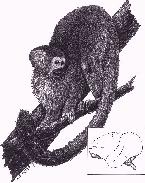
Alfonso Gozalo and Manuel Tantaleán
U.S. Naval Medical Research Institute Detachment and San Marcos
University, Lima, Peru
Introduction
The New World species of primates (Platirrhini) are classified taxonomically according to their anatomical, physiological and behavioral characteristics into three families: Callithrichidae, Cebidae and Callimiconidae (Kavanagh, 1984). Several of the 76 species distributed among these families have been used in biomedical research, but seldom used as pets or for food by indigenous inhabitants of Amazonia. Approximately 13 (40%) of their parasites have been shown to be pathogenic for primates, and 11 species have been associated with human disease (Brack, 1987). Therefore, it is important to be able to identify the species of parasites associated with each of the species of nonhuman primates.
The existing information on protozoa of neotropical primates has been published as scattered reports among several different scientific journals. Consequently, a readily available comprehensive reference source is not available for veterinarians or physicians to rapidly identify the parasites associated with wild captured primates. The purpose of this report is to provide an overall summary of the characteristics of the species of Flagellata, Sarcodina, Sporozoa and Ciliata protozoan parasites associated with neotropical primates, including life cycle, clinical symptoms, pathology, diagnosis, and treatment.
Flagellata
Several species of Flagellata have been reported to affect New World primates (Table 1).
Trypanosoma cruzi. The trypomastigote of this species is found in the blood, adopts the U, C or S form, and measures approximately 20 um long (Figure 1). The central nucleus and large kinetoplast are located in the acute end of the body; it possesses an undulating membrane bordered by a flagellum that is free at one end of the body (Flynn, 1973). This protozoan is the only member of the family Trypanosomatidae that forms amastigote nests in which this stage reproduces. These nests are formed in various tissues of warm-blooded vertebrates. Infection occurs when the metacyclic trypomastigotes, shed in the feces of the arthropod vector, enter the wound caused by the sting of the insect. The vectors are hematophaga insects of the family Rediviidae (Baker, 1972; Flynn, 1973). Unlike other species of trypanosoma, T. cruzi is extremely pathogenic. Experimental infections of nonhuman primates resulted in signs of cardiac insufficiency (edema, bilateral enlargement of the cardiac shadow) and/or death due to myocarditis (Dunn et al., 1963; King, 1976; Toft, 1986). Microscopic diagnosis can be made by observing the parasites in peripheral blood or in histologic sections of cardiac muscle (Flynn, 1973). Drugs of the Imidazole group are recommended for treatment. Fexinidazole is effective against the trypomastigote, but produces anemia and weight loss. Ketoconazole (5-15 mg/k/day) prevents infection by the amastigote during the generalized infection phase, but it must be used with caution because of liver toxicity and inhibition of the synthesis of estheroids from the adrenal glands (Wolff, 1990).
Figures 1-5: Flagellata.
Trypanosoma sanmartini is considered an aberrant strain or subspecies of T. cruzi. This species is usually observed as a curved or S form, with a large ovoid kinetoplast in the margin and moderately developed undulating membrane. The parasite is about 17-24 um long. This species has been found in Saimiri monkeys, but its pathogenicity is unknown (Baker, 1972; Toft, 1986; Wolff, 1990).
Trypanosoma minasense is 29-46 um long; the nucleus is generally located in the middle of the body (Figure 2). The kinetoplast is small and previous to the posterior end. The undulating membrane is well developed and the free flagellum is one-sixth to one-third the length of the body. T. minasense is not pathogenic, but it can be found in mixed infections with T. cruzi (Baker, 1972; Toft, 1986; Wolff, 1990).
Trypanosoma rangeli is larger than T. cruzi, approximately 26-36 um long, with the nucleus located toward the anterior region and kinetoplast small and subterminal (Figure 3). The infective stage is found in the salivary glands of the vector and is inoculated into the host by the sting. The vectors are members of the family Rediviidae. Apparently it is not pathogenic for primates (Baker, 1972; Toft, 1986; Wolff, 1990).
Trypanosoma saimirii and T. diasi are similar in their morphological and biological features, and resemble T. minasense. T. saimirii is 19-29 um long, with a flagellum extending 6-9 um from the end. The kinetoplast is relatively small and is located 5 um from the posterior end. T. diasi is 33-36 um long with a free flagellum extending 7-9 um from the end. Apparently they are not pathogenic for primates (Baker, 1972; Toft, 1986; Wolff, 1990).
Trypanosoma lambrechti is 30-43 um long, including a free flagellum of about 11-12 um (Figure 4). It is easily differentiated from the rest of tripanosoma because of the marked posterior position of the nucleus (Baker, 1972; Toft, 1986; Wolff, 1990).
Diagnosis of all these hemoflagellates is performed by examining blood smears with a light microscope. There are no effective treatment strategies; preventive measures are based on control of the insect vector (Flynn, 1973; Wolff, 1990).
Leishmania brasiliensis: complex members have been reported in Panamanian Saguinus geoffroyi and Aotus trivirgatus (Brack, 1987). These intracellular microorganisms are rounded (1-3 um), ovoid, or elongated (2-4 um by 1.5-2.5 um) (Figure 5). The cytoplasm is homogeneous, but often with minute vacuoles; the nucleus is often large and irregular; the blepharoplast stains more deeply. The number of parasites in a host cell varies from a few to 100 (Kudo, 1977). Oral and nasal mucous membrane ulcerations are observed. Diagnosis is made by observing the parasites microscopically in lesion scrapings (Kudo, 1977). These parasites are transmitted by Phlebotomus and Lutzomyia insects (sandflies), but nonhuman primates are not considered to play an important role in the life cycles of the American Leishmania species (Brack, 1987).
Figures 6-11: Flagellata.
Chilomastix mesnili is a pyriform enteric flagellate whose cystic stage is shaped like a lemon. The trophozoite has a diagonal cleft, nucleus in anterior position, three anterior flagella and a short undulating fourth. It is about 6-24 um long by 3-10 um wide (Figure 6). The cyst is 6.5-10 um long (Figure 7). Infection occurs by ingesting the cyst. It is not pathogenic for primates unless the host is heavily infected (Burrows, 1972; Flynn, 1973; Toft, 1986).
Pentatrichomonas hominis is pyriform, 8-20 um long by 3-14 um wide (Figures 8 and 9). Usually it has five anterior flagella but in some cases can have four or even only three. It is not pathogenic for monkeys (Brady et al., 1988; Burrows, 1972; Flynn, 1973; Toft, 1986).
Trichomonas hominis resembles P. hominis, and is characterized by the presence of 4 free flagella and an undulating membrane bordered by a flagellum that is free posteriorly. It has a vesicle-like nucleus located in the anterior end. There are no known cystic stages. The trophozoite is 5-15 um. Infection of the host is by the oral route (Brady et al., 1988; Burrows, 1972; Flynn, 1973; Toft, 1986).
Giardia lamblia is shaped like a tennis racket without a handle. The trophozoite is 5-15 um long, has two nuclei, 8 flagella (4 lateral, 2 ventral and 2 caudal), and two axostyles (Figure 10). A sucking disc in the anterior ventral face allows the parasite to attach to the intestinal mucosa (Kudo, 1977). The cyst is oval with 4 nuclei, usually located in a pole, axostyles, and remains of flagella (Figure 11). The cyst is the infectious form; infection occurs by the oral route (Kudo, 1977). It is reported to produce diarrhea in monkeys (Burrows, 1972; Flynn, 1973; Toft, 1986). Diagnosis is performed by microscopic observation of the parasites in fecal specimens. Treatment is Metronidazole (35-50 mg/k/day BID for 10 days).
     ------------------------------
           Trypanosoma cruzi
        Trypanosoma sanmartini
         Trypanosoma minasense
          Trypanosoma rangeli
         Trypanosoma saimirii
           Trypanosoma diasi
        Trypanosoma lambrechti
        Leishmania brasiliensis
          Chilomastix mesnili
           Chilomastix spp.
       Pentatrichomonas hominis
          Trichomonas hominis
            Giardia lamblia
              Giardia spp.
     ------------------------------
Table 1: Flagellata parasites reported in neotropical primates.
Sarcodina
The parasites of this group are less frequently observed than Flagellata in neotropical primates. However, several species of intestinal amoebas have been reported, suggesting the possibility of cross-infections in captive primates (Table 2).
Entamoeba histolytica. The trophozoite (Figure 12) measures 15-20 um, but the invasive forms are larger. E. histolytica make progressive movements by means of ectoplasmic pseudopoda. The nucleus has cyst-like form. It presents fine nuclear chromatin, uniformly distributed, and a small endosome, almost always centrally located. The invasive forms can contain red blood cells in their cytoplasm. Precystic uninucleated forms with a great central vacuole surrounded by chromatoid bodies (Figure 13) or pre-cystic forms containing 2 nuclei and rod-shaped chromatoid bodies (Figure 14) are sometimes observed. The mature cyst (Figure 15), which is the infectious form, measures 12-15 um in diameter and has 4 nuclei with the same characteristics as those of the trophozoite (Kudo, 1977; Flynn, 1973). It reproduces by binary fission. The host is infected by ingesting mature, tetranucleated, cysts (Flynn, 1973). Pathogenicity varies with the parasite strain, nutritional condition of the host, environmental factors, and the enteric bacterial flora (Kudo, 1977). Primates of the New World are more susceptible than those of the Old World. Clinically, diarrhea and sometimes dysentery are observed. Chronic colitis, congestion, petechial hemorrhages, and sores occur (Burrows, 1972). The amoebas invade the mucous membrane, forming small colonies that then extend to the submucosa and occasionally to the muscularis mucosae. They produce typical sores in the form of a bottle which vary in size from a few millimeters to large sections of the colon due to the confluence of the lesions. The trophozoites occasionally penetrate to the mesenteric veins and are carried via blood to the liver, lungs, brain, and other organs where they cause abcesses (Flynn, 1973, Toft, 1986, Wolff, 1990). Diagnosis is done by microscopic identification of the organism in the feces or in the lesions. Metronidazole (35-50 mg/k/day BID for 5-10 days) or Paromomicine (25-30 mg/k/day BID for 5-10 days) (Wolff, 1990) are recommended for treatment.
Figures 12-27: Sarcodina.
Entamoeba chattoni. The trophozoite measures 9-25 um and contains only one nucleus with variable characteristics (Figures 16 and 17). The cyst measures 6-18 um and has one nucleus with variable characteristics (Figures 18 and 19). This parasite is found in the cecum and colon. It is not known if it is pathogenic (Burrows, 1972; Flynn, 1973, Toft, 1986).
Iodamoeba butschlii measures 12-15 um; the trophozoite (Figure 20) has a vesicle-like nucleus, without nuclear chromatin and with a large endosome surrounded by refractile granules difficult to visualize. The cyst (Figures 21, 22 and 23), measures 10-12 um. It can be ovoid, ellipsoidal, or triangular with a great well-defined glucogenic vacuole. This stage is the infective form, which enters the host by the oral route. It is found in the cecum and colon. It is not pathogenic (Burrows, 1972; Flynn, 1973, Toft, 1986).
Endolimax nana. A small amoeba, the trophozoite (Figures 24 and 25) measures 2-10 um and the cyst (Figures 26 and 27), 6-8 um. The nucleus lacks chromatin, but the endosome is large and irregular. The cyst, which is the infectious form, is generally tetranucleated. It affects the cecum and colon. It is not pathogenic (Burrows, 1972; Flynn, 1973, Toft, 1986).
Diagnosis of these amoebiases is done by identifying the cysts by fecal smear microscopic examination. The recommended treatment is Metronidazole (35-50 mg/k/day BID for 5-10 days) or Paromomicine (25-30 mg/k/day BID for 5-10 days) (Wolff, 1990).
     ------------------------------
        Entamoeba  histolytica
         Entamoeba  chattoni
         Iodamoeba  butschlii
            Endolimax nana
            Endolimax spp.
     ------------------------------
Table 2: Sarcodina parasites reported in neotropical primates.
Sporozoa
Sarcocystis spp. The cysts are found in skeletal and cardiac muscle. They are cylindrical, ellipsoidal, or irregular in structure. The trophozoites are banana-shaped with the anterior end slightly sharp and the posterior end rounded (Kudo, 1977). They vary in size according to species. An incidence of 16% infection has been reported in callitrichidae at one laboratory (Flynn, 1973). Animals are infected by ingesting trophozoites embedded in muscular tissue or free in the feces (Figure 28). The trophozoites cross the intestinal barrier, enter the blood stream, and migrate to the striated muscle where they are encysted (Figure 29). They divide by binary fission or by schizogonia in trophoblasts surrounded by a cystic wall, continue dividing by binary fission, and then change to trophozoites. As the cyst ages, the trophozoites in the center degenerate and disappear. Once the cyst matures, the wall disintegrates and the trophozoites are released. These migrate through the blood stream to the digestive tract where they are eliminated with the feces (Soulsby, 1987). They are relatively nonpathogenic and do not produce clinical signs. The parasite destroys only the cell it occupies and can produce atrophy by compression of the adjacent cells (Baker, 1972; King, 1976; Toft, 1986). Diagnosis is by microscopic identification of the characteristic cyst (Flynn, 1973). There is no known treatment.
Figures 28-38: Sporozoa.
Cryptosporidium spp. have been reported in the Saimiri monkey (Brack, 1987). It is a small coccidian parasite of poor host specificity. It inhabits the villous border of the enterocyte, mainly of the small intestine. It develops within the microvilliar region but not within the cell. An asexual schizogony is followed by a sexual gametogony; the fertilized macrogametes develop in oocysts 2-6 um in diameter which are eliminated with the feces; they are infectious (Figure 30). The oocysts possess four sporozoites without sporocysts (Melvin & Healy, 1985; Soulsby, 1987). Clinically they produce diarrhea in juvenile or immunodeficient individuals (Acha & Szyfres, 1989). Histologically, neutrophilic infiltration, mainly of the distal portion of the small intestine with thickening, fusion, and flattening of the villiae, as well as necrosis and sloughing of enterocytes, occurs. Similar lesions have been observed occasionally in the walls of the gallbladder duct and pancreatic duct (Brack, 1987). Diagnosis is by observing the microorganisms in the feces through phase-contrast microscopy or special stains (Kynjoun modified acid-fast or Auramine), or in histologic sections of small intestine dyed with Giemsa or Toluidine Blue (Acha & Szyfres, 1989; Brack, 1987; Soulsby, 1987). No treatment is known at present.
Isospora arctopitheci. The oocysts are ellipsoidal, measuring 25.5-30.5 by 23-25.5 um, with smooth walls and no residual body (Figure 31). Sporulation requires 48 hours. The ellipsoidal sporocysts measure 15 by 10 um and contain four elongated sporozoites (Burrows, 1972). The monkey is infected by ingesting oocysts that have been eliminated with the feces; the sporozoites are then freed. They enter the epithelial intestinal cells where the schizont is formed. This contains merozoites that become macro- and microgametocytes that join to form the zygote. The zygote forms a wall, is converted into an oocyst and is eliminated with the feces. They have been reported in wild-caught callitrichids (Burrows, 1972). The infection is extremely rare. Symptoms range from mild diarrhea to dysentery, anorexia, weight loss, eosinophilia, anemia, and, occasionally, death. The ileum is mainly affected, with inflammation and hemorrhage. The mucous membrane is swollen, presenting ulceration and sloughing. Diagnosis is based on recognition of the oocysts in the feces. There is no treatment.
Plasmodium brasilianum is reported as extremely pathogenic in Cebidae and Callitrichidae, producing anemia, fever (quartan malaria), hepatic and splenetic enlargement, depression, and death. This parasite is similar to P. malariae of man. It is thought to be a mutant variety of P. malariae which was introduced by explorers in the Amazon basin region. Schizogony occurs every 72 hours. The schizonts are in the form of a band and the merozoites number 8-10 (Figures 32 and 33). The gametocytes are round and small. The vector is a mosquito of the Anopheles genus (Brack, 1987; Dunn & Lambrecht, 1963; Voller, 1972). Diagnosis is made by observation of the parasite in blood smears with a light microscope. Recommended treatment is Cloroquine phosphate (10 mg/k IM, followed by 5 mg/k at 6 hours, then 5 mg/k/day for two days) and Primaquine (0.3 mg/k/day for 14 days) (Wolff, 1990).
Plasmodium simium has been reported in Alouatta and Brachyteles from Brazil. Schizogony occurs every 48 hours. The schizonts have an amoeboid form; the merozoites number 15-30 (Figures 34 and 35). The gametocytes are large and solid. It is believed that this kind of malaria corresponds to P. ovale or to P. vivax of human origin that has adapted to the monkey (Brack, 1987).
Toxoplasma gondii has been reported as spontaneously infecting Cebidae and Callitrichidae. The definitive hosts are feline; sexual reproduction occurs in the domestic cat. The resulting oocysts (Figure 36) are eliminated with the feces. When they mature, they infect various vertebrates, such as the monkey, by the oral route (Kudo, 1977). The oocysts measure 10-14 um by 10-12 um. They are indistinguishable from those of Isospora bigemina. The infection can also be acquired by eating poorly cooked meat from other intermediary hosts containing cysts of the parasite or, indirectly, congenitally (Flynn, 1973). The extracellular trophozoites (tachizoites) have a banana shape and measure 4-8 um by 2-4 um with a broadened end in which the nucleus is found (Figure 37). The intracellular trophozoites (bradyzoites) vary in form but usually are almost rounded and are found in the cytoplasm of the invaded cell. The cysts are formed in various organ cells, are spherical, and contain a great quantity of bradyzoites (Flynn, 1973; Krogstad et al., 1985; Figure 38). It is believed that primates can get infected in their natural habitat as well as in captivity. The disease is reported to be extremely pathogenic (Brack, 1987; King, 1976; Seibold & Wolf, 1971; Toft, 1986; Wolff, 1990). Clinical signs are anorexia, neurological symptoms, and diarrhea. Diagnosis is made through serology and/or observation of the parasite on histologic sections. Sulfadiazine (120 mg/k), Pirimetamine (1 mg/k) and Clindamicine (10-40 mg/k/day distributed in 3-4 equal doses) are recommended for treatment (Wolff, 1990).
Encephalitozoon cuniculi (=Nosema cuniculi). The trophozoites are straight or slightly bent rod shapes; they measure 2-4 um by 1.2-2.5 um, with an eccentric nucleus. The pseudocysts contain 100 or more trophozoites and are found in nerve cells, macrophages, and other cells. Unlike T. gondii, they do not possess a cystic wall (Flynn, 1973). They multiply by schizogonia. Little is known about the transmission mechanism. Congenital infection occurs. The microorganism is excreted in urine. It usually does not cause clinical symptoms, but encephalitis and nephritis have been observed in the rabbit and dog (Flynn, 1973). It occasionally produces microgranulomas in brain and cerebellum, perivascular cuffing, focal infiltration, and mild meningitis. Granulomas and lymphocytic infiltrations are occasionally observed in the heart and kidney (Flynn, 1973). In primates, its pathogenicity is not known (Baker, 1972; Toft, 1986). Diagnosis is made by microscopic identification of the lesions and the microorganism (Flynn, 1973). No treatment is known.
Haemobartonella spp. Described in Saimiri monkeys in captivity with normocytic and normochromic anemia (Adams et al., 1984). It is classified as an obligated prokaryotic hemotrophic parasite, member of the family Anaplasmataceae, order Rickettsiae. They are spherical organisms 230-330 nm in diameter, present alone or in groups, indented deeply in the plasmatic membrane of the erythrocyte (Adams et al., 1984; Peters et al., 1974). They infect a wide variety of animals. The infections usually are clinically asymptomatic. Diagnosis is made by observation of the microorganisms in blood smears dyed with Giemsa (they are observed as basophilic dottings in the surface of the infected erythrocyte) and confirmed through electron microscopy (Adams et al., 1984). Neither pathogenicity nor treatment is known.
Eperythrozoon spp. have been described in a splenectomized Aotus monkey (Peters et al., 1974). It is similar morphologically to Haemobartonella spp. but, unlike the latter, it is found poorly adhered to the erythrocyte and occasionally free in the plasma (Peters et al., 1974). Diagnosis is accomplished as above. Treatment is not known.
     ------------------------------
         Sarcocystis spp.
       Cryptosporidium spp.
       Isospora arctopitheci
         Isospora callimico
       Plasmodium brasilianum
          Plasmodium simium
          Toxoplasma gondii
       Encephalitozoon cuniculi
         Haemobartonella spp.
          Eperythrozoon spp.
     ------------------------------
Table 3: Sporozoa parasites reported in neotropical primates.
Ciliata
Only one Ciliata species, Balantidium coli, is mentioned in neotropical monkeys in the literature. It affects Alouatta, Ateles and Cebus.
Figures 39-40: Ciliata.
Balantidium coli is the largest protozoan parasite: the trophozoite measures 40-70 um (Figure 39) and the cyst 50-55 microns (Figure 40). The trophozoite is ovoid and covered by cilia, with an attenuated end where the cytostome opens. The cytopyge is found on the opposite end. It has two nuclei, the larger (macronucleus) with a kidney-like form and the smaller (micronucleus) difficult to visualize. The infective form is the cyst, which enters the organism by the oral route (Burrows, 1972; Flynn, 1973). It may or may not be associated with diarrhea. In severe cases it produces ulcerative colitis (Brack, 1987; Toft, 1986). Diagnosis is made through observation by light microscope of the cysts in fresh fecal smears. Metronidazole (35-50 mg/k/BID for 5-10 days) is recommended for treatment (Wolff, 1990).
References
Acha, P. N. & Szyfres, B. (1989). Zoonoses and Communicable Diseases Common to Man and Animals. 2nd Ed. Scientific Publication No. 503. Washington, DC: Pan American Health Organization.
Adams, M. R., Lewis, J. C., & Bullock, B. C. (1984). Hemobartonellosis in Squirrel Monkeys (Saimiri sciureus) in a Domestic Breeding Colony: Case Report and Preliminary Study. Lab Animal Science, 34[1], 82-85.
Baker, J. R. (1972). Protozoa of Tissue and Blood. In R. N. T.-W.-Fiennes, (Ed.) Pathology of Simian Primates, Part II: Infectious and Parasitic Diseases (pp. 29-56). New York: S. Karger.
Brack, M. (1987). Agents Transmissible from Simians to Man. Berlin: Springer-Verlag.
Brady, A. G., Pindak, F. F., Abee, C. R., & Gardner, W. A., Jr. (1988). Enteric Trichomonads of Squirrel Monkeys (Saimiri spp.): Natural Infestation and Treatment. American Journal of Primatology, 14, 65-71.
Burrows, R. B. (1972). Protozoa of the Intestinal Tract. In R. N. T.-W.-Fiennes, (Ed.) Pathology of Simian Primates, Part II: Infectious and Parasitic Diseases (pp. 2-28). New York: S. Karger.
Dunn, F. L. & Lambrecht, F. L. (1963). The hosts of Plasmodium brasilianum, Gonder and von Berenberg-Gossler, 1908. Journal of Parasitology, 49, 316-319.
Dunn, F. L., Lambrecht, F. L., & du Plessis, R. (1963). Trypanosomes of South American monkeys and marmosets. American Journal of Tropical Medicine and Hygiene, 12, 524-534.
Flynn, R. J. (1973). Parasites of Laboratory Animals. Ames, IA: Iowa State University Press.
King, N. W. (1976). Synopsis of the pathology of New World monkeys. In First Inter-American Conference on Conservation and Utilization of American Nonhuman Primates in Biomedical Research (pp. 169-198). Scientific Publication No. 317. Washington, DC: Pan American Health Organization.
Krogstad, D., Visvesvara, G., Walls, K. W., & Smith, J. W. (1985). Blood and Tissue Protozoa. In E. H. Lennette, A. Balows, W. J. Hausler, Jr., & H. J. Shadomy (Eds), Manual of Clinical Microbiology, 4th Ed. (pp. 612-630). Washington, DC: American Society for Microbiology.
Kudo, R. R. (1977). Protozoology, 5th Ed. Springfield, IL: Charles C. Thomas Publishers.
Melvin, D. M. & Healy, G. R. (1985). Intestinal and Urogenital Protozoa. In E. H. Lennette, A. Balows, W. J. Hausler, Jr., & H. J. Shadomy (Eds), Manual of Clinical Microbiology, 4th Ed. (pp. 631-650). Washington, DC: American Society for Microbiology.
Peters, W., Molyneux, D. H., & Howells, R. E. (1974). Eperythrozoon and Haemobartonella in monkeys. Annals of Tropical Medicine and Parasitology, 68, 47-50.
Seibold, H. R. & Wolf, R. H. (1971). Toxoplasmosis in Aotus trivirgatus and Callicebus moloch. Lab Animal Science, 21, 118.
Soulsby, E. J. L. (1987). Parasitología y enfermedades parasitarias en los animales domésticos. 7ma ed., México D.F.: Nueva Editorial Interamericana S.A.
Toft, J. D., II. (1986). The pathoparasitology of nonhuman primates: A review. In K. Benirschke (Ed.) Primates: The Road to Self-Sustaining Populations (pp. 571-679). New York: Springer-Verlag.
Voller, A. (1972). Plasmodium and Hepatocystis. In R. N. T.-W.-Fiennes, (Ed.). Pathology of Simian Primates, Part II: Infectious and Parasitic Diseases (pp. 57-75). New York: S. Karger.
Wolff, P. L. (1990). The Parasites of New World Primates: A Review. Proceedings of the American Association of Zoo Veterinarians, 87-94.
----------------------------------------------------------------------------------
First author's address: Editorial Assistant, NAMRID, Unit Number 3800,
American Embassy, APO AA 34031.
We thank Mr. Ramón Córdova for preparing the illustrations that are presented in this article and Mr. Walter Griebenow for editing the illustrations on the computer.
This study was supported by Walter Reed Army Inst. of Research work unit no. EN241204 63302A .810FH 1531.
The opinions and assertions contained herein are the private ones of the
authors and are not to be construed as official or reflecting the views of the
Dept of the Navy or the Peruvian Government.
----------------------------------------------------------------------------------
* * *
Exequiel M. Patiño, Juan T. Borda, and Julio C. Ruiz
Centro Argentino de Primates (CAPRIM)
Introduction
Cebus apella is a primate with wide distribution in South America. It is found from Colombia and northeastern Brazil to northern Argentina and southeastern Brazil (Brown & Colillas, 1983). In Argentina it lives in the provinces of Misiones, Salta, and Jujuy (Chalukian, 1985).
In the forest, this species lives in social groups composed of one to four adult and subadult males and one to four females of reproductive age with their young. A definite seasonality in its reproductive cycle is seen (Janson, 1984).
In order to establish breeding groups in captivity, starting with wild monkeys of unknown age, we must find parameters that will permit us to determine the monkeys' reproductive capability (Patiño et al., 1981). One of the most important factors to consider with Cebus apella is the age of onset of reproduction, since the majority of wild-caught animals are juveniles (Nagle & Denari, 1982b).
In the present paper we determine criteria for reproductive maturity and document seasonal breeding behavior in Cebus apella born and maintained in outdoor cages.
Subjects, Materials, and Methods
The Primate Center of Argentina (CAPRIM) has a colony of Cebus apella which was founded with 47 monkeys imported from Paraguay during the years 1975-1980. In 1989-1990 more monkeys were added, both from Paraguay and from Misiones Province in Argentina, the majority of them juvenile and infant males. At present (1996) our colony consists of 78 individuals (48 males and 30 females), of which 28 have been born in captivity.
The monkeys are kept in 15m3 outdoor cages, in harems consisting of one male with two to three adult females, or in groups consisting of three to four juveniles or subadults. The monkeys are fed commercial pellets (Cargill ®.) with a minimum protein content of 25%, seasonal fruits, and water ad libitum.
Reproductive maturity was established on the basis of chronological age (true age of those born in captivity, estimated age in the case of the wild-caught monkeys); body weight; testicular volume (V = 3/4(pi)ab2); dentition; pregnancy (determined by manual palpation of the abdomen); and signs of having previously given birth (tearing of the vulva and pigmentation of the mammillae).
In order to determine whether reproduction was seasonal, we recorded the dates of all births between 1989 and 1995.
Results
Figure 1: Body weight and sexual maturity of Cebus apella born at CAPRIM.
Body weight and its relation to sexual maturity: Young born at CAPRIM and weighed immediately after birth (n=10) had body weights averaging 204 +/- 15 g, the males weighing 208 +/- 8 g (n=5), and the females weighing 200 +/- 21 g (n=5).
In females, body weight increased steadily after birth until the age of five years, after which it reached a plateau. In contrast, the males' body weight increased continuously until they were seven years old (see Figure 1).
Three females, all born at CAPRIM, became pregnant for the first time when they were between 6 and 7 years old (Figure 1), with body weights averaging 2017 +/- 293 g, and full dentition (I 2/2; C 1/1; PM 3/3; M 3/3). After their first delivery tearing of the vulva and pigmentation of the mammillae were observed.
The wild-caught females at CAPRIM (n=13) became pregnant when they had body weights averaging 2068 +/- 201 g. The average body weight of both groups of females (captive-born and wild-caught) was 2039 +/- 202 g.
Paternity has been verified in one captive-born male so far: He was 84 months old (Figure 1), with body weight of 3600 g, testicular volume of 4067 mm3, and complete dentition (I 2/2; C 1/1; PM 3/3; M 3/3).
The wild-caught males (n = 4) produced verified pregnancies at CAPRIM with ages estimated at no younger than 7 years, body weights averaging 3775 +/- 179 g, and testicular volumes of 3028 +/- 626 mm3.
These ages and weights for CAPRIM's males and females with verified sexual maturity are similar to those described by Nagle & Denari (1982a) for classifying Cebus apella as fully adult. These authors state that Cebus apella is never sexually mature before 6 or 7 years of age, with weights of 2.0 to 2.8 kg for females and 2.7 to 3.8 kg for males.
Figure 2: Seasonal births of Cebus apella at CAPRIM.
Seasonal reproduction: Between 1989 and 1995, we recorded 36 births, at all times of the year, but with a definite concentration of births (83%) between the months of September and February (that is, in spring and summer) (Figure 2). The reproductive season (the season with the most sexual activity) lasted from March until August, that is, in autumn and winter. This is similar to what Colillas (1986) and Zunino (1990) observed in outdoor cages, and to what has been described for monkeys living in the wild by Janson (1984).
Although Cebus apella does not show seasonal breeding under controlled ambient conditions (i.e., in indoor colonies with lighting, etc., that does not change seasonally) but produces births throughout the year (Nagle & Denari, 1982b), it is a seasonal breeder both in the wild and in captivity when caged outdoors.
References
Brown, A. D. & Colillas, O. J. (1983). Ecología de Cebus apella. A Primatologia No Brasil. Anais 1 deg. Congresso Brasileiro de Primatologia, 301-312. Belo Horizonte, Brasil.
Chalukian, S. C. (1985). Comportamiento Alimentario de Cebus apella Paraguayanus en el Parque Nacional el Rey, Salta. Boletín Primatológico Argentino 3[1], 15-26.
Colillas, O. J. (1986). Biología Reproductiva en Primates Neotropicales. Boletín Primatológico Argentino, 4 [l], 96-117.
Janson, C. H. (1984) Female Choice and Mating System of the Brown Capuchin Monkey Cebus apella (Primates: Cebidae). Zeitschrift für Tierpsychologie, 65, 177-200.
Nagle, C. A. & Denari, J. H. (1982a). The Reproductive Biology of Capuchin Monkeys. International Zoo Yearbook 22, 143-150.
Nagle, C. A. & Denari, J. H. (1982b). The Cebus Monkey (Cebus apella). In J. Hearn (Ed.), Reproduction in New World Primates: New Models in Medical Science (pp. 39-67). Lancaster, England: MTP Press Ltd.
Patiño, E. M., Constantini, M. G., & Claver, J. C. (1981). Evolución de la Capacidad Reproductiva en Monos Cebus apella y Saimiri sciureus. Resúmenes Quintas Jornadas Veterinarias de Corrientes (p. 16). Corrientes, Argentina: Facultad de Ciencias Veterinarias del Universidad Nacional de la Nordeste..
Zunino, G. E. (1990). Reproducción y Mortalidad de Saimiri boliviensis y Cebus apella en Cautiverio. Boletín Primatológico Latinoamericano, 2[1], 23-28.
----------------------------------------------------------------------------------
Authors' address: Centro Argentino de Primates (CAPRIM), Consejo Nacional de
Investigaciones Científicas y Técnicas (CONICET). C. C. 145
(3400) Corrientes, Argentina.
This study was supported in part by Secretaría de Ciencia y
Técnica (SECYT), Univ. Nacional del Nordeste (UNNE).
----------------------------------------------------------------------------------
* * *
Jens Kerl and Hartmut Rothe
Institute of Anthropology, University of Göttingen
Introduction
Laboratory studies on the biology and behavior of the common marmoset (Callithrix jacchus) have sometimes revealed widely different results. For example, Tardif et al. (1986) and Rothe et al. (1993) report that the eldest son of a marmoset family contributes most of all non-reproductive group members to infant-carrying, whereas Box (1977) and Ingram (1977) observed the eldest daughter occupying this position. Furthermore, in the marmoset colony of Johnson et al. (1991), only 33.3% of the adult females got pregnant in the first or second ovarian cycle after being paired, compared to 89.0% of our C. jacchus females (König et al., 1990). And finally, according to Johnson et al. (1991), 11.1% of the infants born to primiparous females have been viable compared to 60.0% in our colony (Rothe et al., 1992).
There are no standards for housing common marmosets in the laboratory, except for a few parameters like room temperature, relative humidity, and the light/dark cycle. In order to quantify the possible influence of some environmental conditions on the physiology and behavior of common marmosets kept under laboratory conditions, an experimental design was developed to investigate the correlation between these parameters and variation in (1) cage size, (2) cage equipment, and (3) group size. A first pilot study, investigating the influence of cage size and cage equipment on the behavior of a pair of common marmosets, was carried out from January to April 1995 by recording the behavior as well as the telemetrically transmitted heart rate (HR) of both animals.
Animals and Methods
The animals were taken from social units of our Callithrix jacchus colony. They were adult at that time (female: 24.1 months; male: 16.3 months), completely socialized, and experienced in infant rearing. HR-transmitters were implanted in their abdominal cavities and they were paired just before the beginning of the pilot study. In order to keep the environment for the animals as stable as possible, the experimental cages were always installed at the same place in the experimental room. In order to reduce stress caused by catching and transporting the animals, they were taught to use a wire-mesh tube to move to a second cage in the neighboring room after each experiment. The three test cages varied in size but were identical in shape (small cage: 1.3 x 1.3 x 1.95 m; medium sized: 1.95 x 1.95 x 2.93 m; large: 2.6 x 2.6 x 3.9 m). Two different sets of cage equipment were used: (1) standard (one feeding shelf, one sleeping box, one (small cage) to eight (large cage) sitting shelves, several fixed and swinging perches); (2) enriched (more than one feeding shelf, more than one sleeping box, fixed perches, natural twigs, wooden screens partially blocking the view so the animals cannot see the entire environment without moving about, free swinging ropes, cage floor covered with a 10 cm layer of woodchips).
Figure 1: Experimental room and home cages. (very large file)
The animals were housed for 12 days under each of the six conditions. In order to measure space utilization, each cage was divided into units of 65 x 65 x 65 cm (small cage: 12 "cells"; middle-sized: 45 cells: large: 96 cells).
The system for collecting behavioral data for up to 10 animals consists of a digitizer-board (DB), with headphones and an electronic mouse-pen, and a personal computer with a serial input. The program allows scan or focal animal sampling and instantaneous or one/zero sampling (Martin & Bateson, 1993).
The 8-channel HR telemetry system enables simultaneous recording from up to eight animals. The single telemetry units (one for each animal) are based on a system that was originally developed and published by Stoehr (1988), modified by adding a demultiplexing unit.
The circuit of the ECG transmitter allows adjustment of the radiated frequency in a wide range of the UHF radio band. A theoretically unlimited number of transmitters can operate simultaneously without interference, per-mitting simultaneous recording of data from all animals in the same cage or enclosure. The range of transmission is 5 m. An operating life of up to 5 months requires a battery of 110 mA-hours' capacity, weighing 1.71g. The weight of the whole transmitter (21.6 15.3 6.1 mm) is 3.14 g. The transmitter is embedded in a beeswax-paraffin mixture and encased in dental acrylic that causes no irritation of the surrounding tissue of the abdomen.
Technical details of signal processing can be obtained from the authors.
Results
Space utilization: Under all conditions the animals, which are arboreal in the wild, spent most of their time in the upper cage. When the floor of the small cage was covered with woodchips the female, but not the male, spent more time in the lowest cells (z-test; p < 0.05) than during other conditions. In the middle-sized and large cages the male, but not the female, spent less time in the lowest cells (both p < 0.01) when the floor was covered with woodchips.
In the standard cages, no correlations were found between the location of equipment (sleeping box, feeding shelf, sitting shelf) and preferred cage cells. In the enriched situation there was a clear preference for cells containing certain items (z-test; p < 0.05).
In the standard cage, the female preferred not to feed at the feeding shelf. Transportable food (offered on five days per week) was always picked up from the feeding shelf and eaten on the opposite side of the cage. In the enriched cages there was always a feeding shelf in the same location as in the standard cage, but it was seldom used. In all enriched cages the animals preferred a sleeping-box located in a different cell than in the standard cages.
Behavior: For both animals, resting behavior was clearly reduced (z-test; p < 0.01), while locomotor (p < 0.01) and exploring (p < 0.01) behavior increased in the enriched cages. Autogrooming and feeding did not change at all. The male's, but not the female's, scent-marking frequency increased in the enriched situation (p < 0.05). Allogrooming was shown more often in the enriched cages by the female (p < 0.01) while it was clearly reduced in the male (p < 0.01).
Heart rate: Mean night heart rate (NHR) was lowered with increasing cage size and seemed to be independent of cage equipment. In cage 6 (large enriched) an unusually high mean NHR (226.44 beats/min-ute) was recorded, though we had expected that it would be the same (167 bpm) as in cage 3 (large standard). However, several copulations were observed in this cage (the female gave birth to twins 5 months later), so this increase of mean NHR was probably influenced by the male's sexual activity. Therefore this result was excluded from the analysis.
The mean daytime heart rate (DHR) increased with increasing cage size for the standard cages. This was expected because of the increased options for locomotor behavior. Most interesting, however, is the complete lack of any changes of mean DHR between the largest standard cage and the smallest enriched cage. From this we conclude that the enriched small cage (cage 4) apparently elicits as much activity as the eight-times-larger standard cage (cage 3). Enlarging the enriched cage volume had no effect on the mean DHR.
Because each animal has a minimum HR (during sleep), the effect of environmental change should best be measurable when this minimum HR is eliminated from the calculations. Differences were calculated between the mean day HR and the mean night HR (DDN = DHR minus NHR, see Figure 8) for each condition. In cage 1 (small standard) a small DDN was found, the result of low activity by day and a rather high NHR. Due to a higher DHR and a lower NHR the DDN was greater in cage 2 (middle-sized standard; 58.59 bpm). The largest DDN (76.52 bpm) was recorded for cage 3 (large standard), in which the NHR was lowest and the DHR was highest, apparently due to high activity.
In cage 4 (small enriched) a high DHR and a high NHR led to a DDN that was smaller than that in cage 3, but still twice that for the same-sized standard cage 1 (61.49 bpm). In cage 5 (middle-sized enriched) the DDN was also higher than that in cage 2 (68.98 bpm). Apparently due to the high sexual activity there was nearly no difference between the DHR and NHR in cage 6 (large enriched). In summary, DDN is a good measure of the effects on HR of combinations of cage size and equipment.
Figure 2: Differences between day- and night-heart rates under each of 6 conditions.
Discussion
Space utilization: The results of this study, although limited by the small number of subjects, allow three important conclusions about the housing of common marmosets in the laboratory: 1) The attractiveness of any given cage equipment (sleeping box, feeding-shelf, resting-shelf) is largely dependent on the rest of the environment inside and outside of the cage and cannot be considered a constant.
2) The place where the feeding-device is mounted may not be where the animals prefer to feed. The feeding behavior of the female showed that the randomly chosen site for installing a single feeding-shelf in the standard cage was not acceptable to her. For adequate housing of marmosets one should test the best site for installing a feeding device. One can offer several sites, letting the animals choose their favorite. When a preferred site is found the additional devices can be removed. Since the preferred location may change over time, perhaps due to the influence of neighboring animals or seasonal changes, this test should be repeated regularly.
3) The place where the animals prefer to rest may not be the location where the sleeping-box is mounted. Caine et al. (1992) have shown for Saguinus labiatus a clear preference for the highest point of the cage. They also tested different types of sleeping boxes and observed a preference for a completely closed type with only one entrance-hole. In the standard cages of our study the single sleeping box was fastened in the middle of one side of the cage. In our enriched cages identical boxes were installed in the same position; additional boxes were installed on the cage sides near the window and the entrance of the room, respectively. The animals showed a clear preference for those sleeping-boxes from which the room entrance could be observed. Thus, the best place for the sleeping-box should also be tested empirically.
Behavior: In agreement with data provided by Chamove (1989) for Callitrichids and by Molzen & French (1989) for Leontopithecus rosalia, our animals rested less (male: 33.0% -> 24.3%) and explored more (male: 1.9% -> 6.9%) when kept in the enriched cages.
From the increasing locomotor and exploring behavior in the enriched cages one might conclude that the observed shift in the time budget is towards a more "natural" one, since in most field reports the proportion of locomotion was much higher than those of the animals in this study. On the other hand, the decrease in resting may imply a shift to a less natural time-budget. When comparing these situations it should be clear what is meant by the term "resting". If it means only "the animal does not move," then even periods of alertness would be included. Schnell (in press) observed a distinct reduction in locomotor behavior--even immobility--as well as tremendous stress tachycardia of about 450 bpm and a hypertonic arterial blood pressure (both recorded by telemetry) in his captive C. jacchus when an animal keeper with catching-gloves entered the room. In this case "resting" would include motionlessness but not relaxing.
From the rather good agreement among captive studies one can conclude that the captive environment produces a behavioral response that cannot be compared to the field situation. Perhaps the lack of resting is an effect of the captive situation itself (high population density) which cannot be overcome by offering larger or enriched cages.
Heartrate: The telemetric measurement of HR turned out to be a powerful tool for detecting the influence of environmental change on the behavior of common marmosets. Stoehr (1988) reported on inanimate influences on the HR of Tupaia belangeri and (1986) revealed persistent influences of the social environment on the HR of these animals by long-term HR-telemetry. Schnell (in press) has observed shifts in the mean HR of Callithrix jacchus, depending on day of the week. Monday to Friday the telemetrically measured HRs were at a stable mean level, while they were lower on the weekend. These examples show how HR-telemetry enables researchers to observe physiological responses of the undisturbed animal that cannot be detected in a handled animal.
The data of this pilot study demonstrate that it is necessary to collect HR-data during day and night. The mean HR by day probably reflects the amount of activity of the animal. In this study it increased with increasing cage size in standard cages and remained stable in all enriched cages. This result can be interpreted as an indication that a sound amount of activity is reached by offering enough space and/or complexity of environment. The NHR responded only to the parameter of cage size (physical space) and not to cage equipment (psychological space, sensu Chamove, 1989).
The difference between the DHR and NHR is a sensitive tool for measuring effects of the inanimate environment. With the exception of Condition 6 (large cage, enriched equipment) there is an HR-difference for the combination of each of the two tested parameters. Assuming that the field situation represents optimum psychological well-being for animals, our results lead to the conclusion that the effectiveness of attempts to enrich the environment of the animals can be determined by the difference between the DHR and NHR. Furthermore, this result indicates clear interdependencies between the inanimate environment and the physiological and behavioral response of the animals. In the future, we may be able to better compare results obtained under different housing conditions (as described in the Materials and Methods sections of published papers) if the mean NHR and the difference between the DHR and the NHR are included.
References
Box, H. O. (1977). Quantitative data on the carrying of young captive monkey (Callithrix jacchus) by other members of their family groups. Primates 18, 475-484.
Chamove, A. S. (1989). Environmental enrichment: A review. Animal Technology 40[3], 155-178.
Caine, N. G., Potter, M. P, & Mayer, E. (1992). Sleeping site selection by captive tamarins (Saguinus labiatus). Ethology 90[1], 63 ff.
Ingram, J. C. (1977). Interactions between parents and infants, and the development of independence in the common marmoset. Animal Behaviour, 25, 811-827.
Johnson, E. O., Kamilaris, T. C., Carter, S., Gold, P. W. & Chrousos, G. P. (1991). "Environmental stress" and reproductive success in the common marmoset (Callithrix jacchus jacchus). American Journal of Primatology, 25, 191-201.
König, A, Radespiel, U., Siess, M., Rothe, H., & Darms, K. (1990). Analysis of pairing, parturition, and interbirth-intervals in a colony of common marmosets (Callithrix jacchus). Zeitschrift für Säugetierkunde, 55, 308-314.
Martin, P. & Bateson, P. (1993). Measuring Behaviour. 2nd ed. Cambridge: Cambridge University Press.
Molzen, E. M. & French, J. A. (1989). The problem of foraging in captive callitrichid primates: Behavioural time budgets and foraging skills. pp. 89-101 In E. F. Segal (Ed.), Housing, Care, and Psychological Well- being of Captive and Laboratory Primates. Park Ridge, NJ: Noyes Publications.
Rothe, H., Darms, K., & König, A. (1992). Sex ratio and mortality in a laboratory colony of the common marmoset (Callithrix jacchus). Laboratory Animals, 26, 88-99.
Rothe, H., Darms, K., König, A., Radespiel, U., & Jünemann, B. (1993). Long-term study of infant-carrying behavior in captive common marmosets (Callithrix jacchus): Effect of nonreproductive helpers on parents' carrying performance. International Journal of Primatology, 14, 79-93.
Schnell, C. (in press) Marmoset telemetry: present applications and future highlights. In L. Scott & C. Price (eds.), Proceedings of the EUPREN/EMRG Winter Workshop 1995, 6-8 Dezember, Göttingen.
Stoehr, W. (1986). Heart rate of tree shrews and its persistent modification by social contact. In T. H. Schmidt, T. M. Dembrowski, & G. Bluemchen (Eds.). Biological and Psychological Factors in Cardiovascular Disease. Berlin und Heidelberg: Springer Verlag.
Stoehr, W. (1988). Longterm heartrate telemetry in small mammals: A comprehensive approach as a prerequisite for valid results. Physiology & Behavior 43, 567-576.
Tardif, S. D., Carson, R. L. & Gangaware, B. L. (1986) Comparison of infant care in family groups of the common marmoset (Callithrix jacchus) and the cotton-top tamarin (Saguinus oedipus). American Journal of Primatology, 11, 103-110.
----------------------------------------------------------------------------------
Authors' address: Institute of Anthropology, University of Göttingen,
Ethologische Station Sennickerode, 37130 Gleichen, Germany [e-mail:
[email protected]].
We wish to thank the following persons: Wolf Stoehr (Bayreuth) for his help in transmitter development, signal processor-construction, and surgical technique. Christian Schnell (Basel) for implantation of the transmitters and helpful hints for the treatment of implanted animals. The veterinarians of the German Primate Center (DPZ, Göttingen) for their help in the care of the implanted animals. Manfred Glahe, Hans Badstuebner and Ullrich Conrad (Göttingen) for their great help in hardware development. Klaus and Ralf Utermoehlen (Göttingen) for their help in construction of the antenna. Kerstin Schibat (Göttingen) for the provision of dental tools that turned out to be a prerequisite for the construction of the transmitters. Fa. Siegert (Cadolzburg) and Fa. Roederstein (Landshut) for generous delivery of samples of miniature electronic components.
Supported by a grant to Ro (DFG Ro 356/14-1).
----------------------------------------------------------------------------------
* * *
Two cynomolgus monkeys were found positive for filovirus at HRP, Inc. in Alice, TX. The monkeys, part of a shipment of 100, were imported on March 21, 1996. They had been in quarantine since arrival. The origin of the monkeys was the Philippines, and they were reportedly colony-raised. The supplier, Ferlite Scientific Research, Inc., was the source of the monkeys in the previous episode of Reston filovirus at the Texas Primate Center in 1990 and at the Reston facility in 1989. There have been no other shipments from this supplier to the United States in 1996.
On March 27, one monkey showed signs of illness, and died on March 30. Necropsy revealed a pneumonic process, and the liver was positive for Reston filovirus by antigen capture. On April 10, a second animal became febrile and "off feed." Serum was found positive for Reston filovirus on April 13 and the animal was sacrificed. As of April 17, no other monkeys had shown signs of illness. Preliminary tests showed that the genetic sequence of the virus was "much more than 90 percent" identical with the Reston strain from the 1988 outbreak.
The second monkey to become ill was housed in a cage at the opposite end of a block of about 25 cages from the index case. Monkeys in the cages adjacent to the ill monkeys were also tested for Reston filovirus.
Routine protective measures for the staff include Tyvek gowns, boots, gloves, dust-mist respirators, and face shields. There has been no unexplained illness or fever among the staff who had contact with the monkeys or specimens (two veterinarians, five handlers and one lab technician). Baseline serum had been banked for these staff members. Two employees had been shown to be seropositive to Reston in a 1993 survey.
Ebola, another filovirus, is one of the world's deadliest diseases, causing 80 percent of its victims to bleed to death. It is spread through bodily fluids, commonly, but not always, through a break in the skin. It has no treatment and no cure. But related filoviruses seem less deadly to humans. The one that struck the Reston importer in 1989, killing dozens of monkeys, is one such strain. Four people were known to have been exposed to the Reston virus, but none became ill.
Dr. Manuel Dayrit, assistant secretary of the Philippine Department of Health, said that Philippine authorities are eager to stop the spread of the disease because Philippine monkeys are used extensively in medical research. The government banned the export of monkeys in April, pending results of an investigation.
On June 10 the Philippines lifted its export ban on four of five monkey breeding farms after tests showed the four were not infected with any strain of the deadly filovirus. Environment Secretary Victor Ramos said monkeys at the fifth facility, Ferlite Scientific Research, Inc., were found to have the "Reston strain" of the virus, and it is still banned from exporting the animals.
Ramos said he had lifted the ban on the four monkey breeding farms after tests showed their animals were not infected. But he said the Departments of Health and Agriculture still must give their approval before any monkeys may be exported.
About 15,000 monkeys intended for export are kept in the five facilities, including 1,600 at Ferlite.
The Ferlite Scientific Research, Inc. monkey farm is a 2.5-3-hectare area in Calamba, Laguna, about 40 km. south of Manila. They have open cages as their holding facilities; the quarantine facility consists of individual cages. In 1995 they exported monkeys to the USA and Sweden. In 1996 Ferlite exported the 100 monkeys to Hazleton in Texas. A second batch of 100 monkeys to be exported to the same facility is still in Ferlite's quarantine facility. Their monkeys are quarantined 30 days prior to shipment. The last replacement of breeders was in November, 1995.
----------------------------------------------------------------------------------
This report was compiled from material published and/or posted to electronic
sources by NABR, WHO, the Federation of American Scientists, and the Texas
Department of Health.
----------------------------------------------------------------------------------
* * *
Veterinary Case Reports
David Lee-Parritz, of the New England RPRC, will be coordinating the case report sessions at the Association of Primate Veterinarians meeting this year. The meeting will be held in Madison, WI in conjunction with the American Society of Primatologists, August 16-18.
He writes: "Case reports have always been a major feature of our clinical meetings of the `monkey doctors'. All attendees are invited to prepare brief presentations of interesting or puzzling cases. Definitive diagnoses are not required! Please look through your records and think of one or two cases to discuss."
Send your titles to David Lee-Parritz, New England RPRC, One Pine Hill Dr., Southborough, MA 01772 [e-mail: [email protected]].
Pan Africa News
The Editors of Pan Africa News gather information from any person working in research and conservation of Pan species (chimps and bonobos) in Africa. Specifically, they welcome information on:
Grateful Med on the Internet
The National Library of Medicine's Grateful Med electronic retrieval service is moving to the Internet, making their storehouse of electronic databases available via the Web. The service, dubbed Internet Grateful Med, does not require any special software, and will be priced per character shipped, with a typical physician's search costing about $1.25. Would-be users need to sign up for the service and receive a user-ID code and a password [http://igm.nlm.nih.gov/ or 800-638-8480]. -- From the Chronicle of Higher Education, 26 Apr 96, A25
Primate Species in Protected Areas
The Neotropical Section of the IUCN/SSC Primate Specialist Group is setting up a data base of the primate species and subspecies living in protected areas. This information is most important for a better understanding of their conservation status. They will be most grateful for any information, unpublished and published, on the occurrence of primates in parks and reserves, and any information on their status in these areas and the status of the protected areas. Their working list of protected areas is based on the IUCN listing published in 1992, and that for the primates on the species list published in the supplement edition of volume 4 of Neotropical Primates, the PSG Neotropical Section newsletter, published in September, 1994.
The information obtained will be fully documented, sources acknowledged, and, hopefully, published in a supplement edition of Neotropical Primates. Any comments regarding either of these lists are also very welcome. "Thank you for helping in this endeavour." Please send information to: Anthony B. Rylands, c/o Conservation International do Brasil, Avenida Antônio Abrahão Caram 820/302, 31275-000 Belo Horizonte, Minas Gerais, Brazil [Tel/Fax: +55 31 441-1795; e-mail: [email protected] or [email protected]].
"Evaluating Eden"
The International Institute for Environment and Development has initiated a three-year research program to investigate and evaluate the environmental, social, and economic dimensions and impacts of community wildlife management (CWM) initiatives in developed and developing countries, and examine the conditions which contribute to successful CWM.
At this stage, they are seeking to identify institutions and individuals who are:
NIH Grants Information e-mail Address
The NIH Grants Information Office, formerly with the Division of Research Grants and now a component of the Extramural Outreach and Information Resources Office, Office of Extramural Research, Office of the Director, NIH, has changed its e-mail address. The new e-mail address is: [email protected] Use this address when requesting single copies of grant application materials or program guidelines and for general questions regarding extramural grant programs. The grants information telephone and FAX numbers remain unchanged. Grant applications and other printed materials may be requested on (301) 435-0714 or by FAX on (301) 480- 0525.
More WWW URLs
The Caribbean Primate Research Center:
ourworld.compuserve.com/homepages/oceanpkvetclin/homepage.htm
Southwest Fnd. for Biomed. Research:
www.sfbr.org/
"NetVet Links," a periodic newsletter summary of new veterinary
websites:
netvet.wustl.edu/whatsnew.htm
German Mountain Gorilla & Rainforest Direct Aid, including
English version of Gorilla Journal:
www.kilimanjaro.com/gorilla/brd
Americans for Medical Progress:
www.ampef.org
The Monkey Sanctuary, Cornwall, U.K.:
ourworld.compuserve.com/homepages/monkey_sanctuary_uk/
A taxonomic hierarchy of all organisms:
www.vet.ed.ac.uk
UC Santa Cruz Physical Anthropology and Archaeology Labs:
zzyx.ucsc.edu/~jjosh/lab.html
Animal Behavior and Welfare sites:
www.wam.umd.edu/~jaguar/welcome.html
New England Journal of Medicine:
www.nejm.org/
REAC:
www.aphis.usda.gov/reac
AALAS:
www.aalas.org
Pan Africa News:
jinrui.zool.kyoto-u.ac.jp/PAN/home.html
Outbreak: the Emerging Disease web site:
www.objarts.com/outbreak/
OPRR :
www.nih.gov:80/grants/oprr/oprr.htm
* * *
Measuring Behavior `96
Measuring Behavior `96, an international workshop on methods and techniques in behavioral research, will be held 16-18 October 1996 in Utrecht, The Netherlands. The meeting is co-organized by Utrecht University and Noldus Information Technology, manufacturer of software and instrumentation for behavioral research.
Presentations will be grouped in three main areas:
The registration fee, which includes lunches and refreshments, is NLG 200 (students: NLG 50) before 1 August 1996, and NLG 300 (students: NLG 75) after that date. Those who cannot afford the registration fee may present a motivated request for a reduced fee to the Local Organizing Committee at the address below. For program booklet, registration/abstract forms, and more information, contact Measuring Behavior `96, Workshop Secretariat, Attn: Rosan Nikkelen, P.O. Box 268, 6700 AG Wageningen, The Netherlands [+31-(0)317-497677; fax: +31-(0)317-424496; e-mail: [email protected]; on the WWW: http://www.diva.nl/noldus/mb96.html].
Animal Care and Use Programs
The NIH Office for Protection from Research Risks (OPRR) sponsors workshops on implementing the Public Health Service Policy on Humane Care and Use of Laboratory Animals. The workshops are open to institutional administrators, members of Institutional Animal Care and Use Committees, laboratory animal veterinarians, investigators, and other institutional staff who have responsibility for high-quality management of sound institutional animal care and use programs. Ample opportunities will be provided to exchange ideas and interests through question-and-answer sessions and information discussions.
A workshop titled "The 1996 Guide for the Care and Use of Laboratory Animals: The Era of Performance Based Standards" will be held September 19-20, 1996 in Denver, CO, cosponsored by the University of Colorado Health Sciences Center and the University of Southern Colorado. There is a $175 registration fee.
For registration, contact Ms. Joann Bauer, Senior Conference Coordinator, Continuing Med. Education Office, Univ. of Colorado Health Sci. Center, Campus Box C295, 4200 East Ninth Ave, Denver, CO 80262 [303-372-9054; 1-800-882-9153; fax: 303-372-9065].
For information concerning future NIH/OPRR Animal Welfare Education Workshops, contact Ms Darlene M. Ross, OPRR, NIH, 6100 Executive Blvd, Suite 3B01, MSC 7507, Rockville, MD 20892-7507 [301-496-8101, Ext. 233; fax: 301-402-0527].
* * *
The Ebola virus is one of the most pathogenic viruses known to science, causing death in 50-90% of all clinically ill cases. Ebola hemorrhagic fever is often characterized by the sudden onset of fever, weakness, muscle pain, headache and sore throat. This is followed by vomiting, diarrhea, rash, limited kidney and liver functions, and both internal and external bleeding. Several different forms of Ebola virus have been identified and may be associated with other clinical expressions, on which further research is required. The incubation period is 2 to 21 days.
Specialized laboratory tests (which are not commercially available) on blood specimens detect specific antigens or antibodies and/or isolate the virus. These tests present an extreme biohazard and are only conducted under maximum containment conditions. No specific treatment or vaccine exists for Ebola hemorrhagic fever. Severe cases require intensive supportive care, as patients are frequently dehydrated and in need of intravenous fluids. Experimental studies involving the use of hyperimmune sera on animals demonstrated no long-term protection against the disease after interruption of therapy.
The Ebola virus was first identified in a western equatorial province of Sudan and in a nearby region of Zaire in 1976. An isolated case occurred in Tandala, Zaire in 1977, a second outbreak occurred in Sudan in 1979, and an epidemic in the Bandundu Region of Zaire in 1995 caused 245 deaths. Two isolated cases of Ebola hemorrhagic fever were also identified in Côte d'Ivoire in 1994-95. The most recent outbreak was in rural Gabon in February, 1996.
The natural reservoir of the Ebola virus is not known. Extensive ecological studies are currently underway in Côte d'Ivoire, Gabon, and Zaire to identify the reservoir. Ebola-related filoviruses were isolated from cynomolgus monkeys (Macaca fascicularis) imported into the United States of America from the Philippines in 1989. A number of the monkeys died and at least four persons were infected, although none of them suffered clinical illness.
The Ebola virus is transmitted by direct contact with the blood, secretions, organs or semen of infected persons. Transmission through semen may occur up to 7 weeks after clinical recovery, as with Marburg hemorrhagic fever. Transmission of the Ebola virus has also occurred by handling ill or dead infected chimpanzees, as was recently documented in Côte d Ivoire. Health care workers have frequently been infected while attending patients. In the 1976 epidemic in Zaire, every Ebola case caused by contaminated syringes and needles died.
Suspected cases should be isolated from other patients and strict barrier nursing techniques practiced. All hospital personnel should be briefed on the nature of the disease and its routes of transmission. Particular emphasis should be placed on ensuring that high-risk procedures such as the placing of intravenous lines and the handling of blood, secretions, catheters and suction devices are done under barrier nursing conditions. Hospital staff should have individual gowns, gloves and masks. Gloves and masks must not be reused unless disinfected. Patients who die from the disease should be promptly buried or cremated.
As the primary mode of person-to-person transmission is contact with contaminated blood, secretions, or body fluids, any person who has had close physical contact with patients should be kept under strict surveillance, i.e. body temperature checks twice a day, with immediate hospitalization and strict isolation recommended in case of temperatures above 38.3deg. C (101deg. F). Casual contacts should be placed on alert and asked to report any fever. Surveillance of suspected cases should continue for three weeks after the date of their last contact. Hospital personnel who come into close contact with patients or contaminated materials without barrier nursing attire must be considered exposed and put under close supervised surveillance.
For more information, please contact the office of Health Communications and Public Relations, WHO Geneva, [4122 791 2584/3223; fax: 791 4858. e-mail: [email protected]]. -- WHO Fact Sheet 103 (Revised) February 1996
* * *
Revision of Guide Complete
The Institute of Laboratory Animal Resources (ILAR) committee charged with revising the Guide for the Care and Use of Laboratory Animals (Guide) has completed its work. A respected resource for decades, the Guide has been revised by a panel of experts, based on input from scientists and the public. The Guide incorporates recent research on commonly used species, including farm animals, and includes extensive references. It is organized around major components of animal use:
The Guide provides a framework for the judgments required in the management of animal facilities. This revision will be important to researchers, animal care technicians, policymakers involved in research issues, and animal welfare advocates.
For information about availability of the revised edition of the Guide contact ILAR, 2101 Constitution Ave, NW, Washington, DC 20417 [202-334-2590; fax: 202-334-1687; e-mail: [email protected]]. Meanwhile, the complete text of the 1985 edition is available at http:/ /netvet.wustl.edu/org/awic/law/phs/guide.txt on the World Wide Web.
Ebola Hemorrhagic Fever, Gabon
No new cases of Ebola hemorrhagic fever have been reported in Gabon since the death of the last case on 12 March 1996. The outbreak was therefore officially declared over on 23 April 1996, after a lapse of 42 days, corresponding to twice the maximum incubation period. The outbreak occurred in the village of Mayibout II, Makokou Health District, Ogooue-Ivindo Province. It was linked to the butchering, transport, and preparation for consumption of a chimpanzee found dead in the forest on 24 January 1996. The total number of cases was 37 (20 males, 17 females) and the mean age was 27 years (range 7 months to 70 years). Rapid identification of the disease and appropriate control measures quickly brought the outbreak under control. -- From Communicable Disease News, 29 April 1996, WHO/EMC
Animal Import News
At the Council of Europe hearing on the transportation of laboratory animals held in Strasbourg on 2 April 1996, representatives from the International Airline Transportation Association (IATA) confirmed that strict interpretation of international aviation law says that airlines are legally obligated to carry all consignments into or out of the airlines' country of origin if asked to do so. They further said that, with regard to strict enforcement for laboratory primates, IATA had an "official" neutral position. As such, the German government has forced Lufthansa to resume carrying primates for laboratory purposes. Primates going to or leaving Germany must be carried by Lufthansa if requested; however the airline could not be forced to carry primates between third countries. This applies to the United States as well--all U.S. airlines could be required to carry animals into and out of the U.S. -- A posting by Jacquie Calnan, Americans for Medical Progress, to CompMed
DPZ Directors
Prof. Dr. Hans-Jörg Kuhn, who was the scientific director of the German Primate Center (DPZ) from its founding in 1977, retired from the directorship February 29, 1996. Even before founding the DPZ he had been the main promoter of the idea of a national primate center in Germany. The institute, with its primate-keeping facilities, laboratories, and offices, was built at the campus of the University of Göttingen between 1979 to 1984. About 200 people work at the DPZ, about 70 of them scientists, in the departments of virology and immunology, reproductive biology, neurobiology, and pathology, and the research groups of ethology, biocommunication, and experimental pathology. The DPZ keeps about 1000 primates of ten species. Prof. Dr. Kuhn was honored in a public ceremony on March 21.
The new scientific director of the DPZ is Prof. Dr. Gerhard Hunsmann, who received his Ph.D. at the University of Würzburg in 1971. He has worked at the Max-Planck-Institut für Virusforschung in Tübingen, the Max-Planck-Institut für Immunbiologie in Freiburg, and the Institut für Immunbiologie at the University of Freiburg. He has been head of the department of virology and immunology at the DPZ since 1983. His main interests are AIDS research, hepatitis research, and prion diseases. A new department of genetics is planned, which will enlarge the scientific spectrum of the center. -- From a posting by Dr. Dr. Michael Schwibbe to Primate-talk
Chimp Josie on the Mend
June 13, 1996--Josephine, the chimp at the Johannesburg Zoo who underwent a ground-breaking heart operation recently, is doing "exceptionally well" and is expected to be reintroduced to her troop next week. Zoo curator Jaqui Thompson said "Josie", believed to be the first chimp in the world to have heart surgery, had been transferred from her small "squeeze" cage to one of the zoo's hospital wards. "The only problem is that she has become very lazy," Thompson said. "She has grown used to being fed by hand. Now, when food is put in front of her, she just looks at it and expects somebody to put it in her mouth." Josie, a grandmother in her 40s and something of an elder in her primate enclosure, had a diseased section of her aorta removed in a four-hour operation last Monday. The section was replaced with a Gortex graft. Thompson said Josie's keepers were "holding thumbs" that the chimp's reintroduction to her troop would go well. "They could reject her, but she's older, so she has that on her side." - - From the Johannesburg Star, posted to Primate-Talk by Greg Hofmeyr, University of Pretoria
New Vets at Primate Foundation of AZ
Jo Fritz has announced that Kathleen Hoffman, D.V.M. (recently at U.C. San Francisco) and Robert Hoffman, D.V.M. (recently of the San Francisco Humane Society) have accepted the position of Staff Veterinarian at the Primate Foundation of Arizona on a "time share" basis. Nominally, "Dr. Kit" will be Chief Veterinarian and "Dr. Rob" Assistant Veterinarian, but they will each work 20 hours/week.* * *
Small-eared Bushbabies Available
Jeannette Ward has small-eared bushbabies (Otolemur garnettii) for sale to research laboratories or established zoological parks. A no-resale contract will be required. No dealers, wholesale or retail. The animals are all captive-born in her laboratory and have been subjects in behavioral research only; no drugs or other invasive procedures. Contact Jeanette at the Psychology Dept, Univ. of Memphis, Memphis, TN 38152 [901-678-2375; e-mail: [email protected]].
Natural History Book Resources
Watkins Natural History Books has just published their Catalogue No. 75, which lists more than 500 used and out-of-print books. The list includes many books in mammalogy, especially mammalian behavior, taxonomy, and ecology. There are also books relating to birds, reptiles, husbandry, and wildlife management. For a copy of this catalogue, contact Larry C. Watkins, Watkins Natural History Books, 7036 State Highway 29, Dolgeville, NY 13329 [518-568-2280].
Books From Bree, owned by Morgan and Shoshana Edwards, specializes in out-of-print books in all of the sciences, including natural history and its related subjects. They will search for any book, in or out of print, at no charge for the search. Contact them at 7795 SW Hall Blvd., Beaverton, OR 97008 [503-644-7218; 1-800-884-0993; e-mail: [email protected]; web site: http://www.auldbooks.com/].
Dissection Alternatives
The Humane Society of the United States (HSUS) has established a loan program to provide students and educators with up-to-date alternatives to classroom animal dissection and live animal experimentation. For a listing of resources available at educational levels from kindergarten through college, including CD-ROMs, computer diskettes, models, videotapes, and charts, contact Jonathan Balcombe, Ph.D., Associate Director for Education, Animal Research Issues, HSUS, 2100 L Street, NW, Washington, DC 20037 [ 301-258-3046; fax: 301-258-3082; e-mail: [email protected]].
Callithrix jacchus Family Available
The Ethologische Station Sennickerode is offering a young family of Callithrix jacchus. The parents have been in a research project (described on pp. 10-14) which combined recording of behavioral and heart rate data and have therefore undergone two surgical procedures (implanting and removing an ECG-transmitter in the stomach cavity). The birth dates are: female 16.12.92, male 11.08.93, both sons 15.09.95. They should now go to a facility where ethological, rather than biomedical, research is done. The family will be given free but shipping costs must be paid by the new owners.
Interested persons should contact Jens Kerl, Ethologische Station Sennickerode, Sennickerode 11, 37130 Gleichen, Germany [+49-5592-1513; fax: +49-5592-1524; e-mail: [email protected]].
* * *
Rwanda Rangers Win Getty Conservation Prize
For risking their lives to stay at their posts and guard endangered mountain gorillas during the 1994 Rwandan civil war, the staff of Rwanda's Parc National des Volcans will receive the J. Paul Getty Wildlife Conservation Award. The $50,000 prize, administered by World Wildlife Fund, is one of the largest awards given for outstanding achievement in the conservation of wildlife and its habitats.
WWF President Kathryn S. Fuller announced the winner in late January: "The past eighteen months have been a time of unspeakable tragedy for the people of Rwanda. The Parc National des Volcans staff, through their heroic efforts, have helped prevent a compounding of that tragedy while protecting mountain gorillas."
The park's troubles began when soldiers breached the park's perimeters and ransacked and damaged its buildings. Later, when remnants of the deposed government's army fled the country, they beat a destructive path straight through the park, breaking into offices, destroying records and books, and throwing computers out windows.
Eventually, the deposed government's army, in an effort to clear out as much of Rwanda's population as possible, forcibly drove park staff into Zaire. When the new Rwandan government finally gained access to the park, they found a heap of buildings and infrastructure, all of it needing to be rebuilt.
The International Gorilla Conservation Program, supported by a coalition of conservation groups, is currently helping to rebuild the park, supplying the Rwandan government with copies of destroyed files, and working with officials to strengthen the country's park system.
Although some of the original staff have since returned to the park, the tourist revenues that once supported it have disappeared, and the park's needs remain enormous. The Getty Prize money will help defray some expenses, while at the same time honoring a group of courageous conservationists as they resume their task of protecting mountain gorillas and their forest habitat. --from WWF Focus Newsletter, March/April 1996, 18[2].
Rozmiarek Honored
Dr. Harry Rozmiarek of the University of Pennsylvania is being awarded the Charles River Prize by the AVMA for distinguished contributions to the field of science and laboratory animal medicine. The award ceremony will be on July 20th at the AVMA meeting in Louisville, Kentucky.
Replacement/Refinement Awards
The Dörenkamp-Zbinden Foundation of Switzerland has announced its awards policy from 1996 on. The Foundation intends making two awards annually, each between 25-50,000 SF. One award will be for research demonstrating in-vitro methods or ethically acceptable experiments in man which can replace the use of animals in experiments. The second award will be made for techniques, instruments or drugs which have produced a clear reduction in suffering in animals used in experiments.
Applications will be judged by a board consisting both of research scientists and lay persons, and will be assessed both for scientific quality and relevance to animal welfare. Nominations for the award (6 copies) should be made to: Prof. Dr. med. Dr. h.c. K. Brune, Institute of Experimental and Clinical Pharmacology and Toxicology, Universtatsstr. 22., D-91054 Erlangen, Germany. The closing date for nominations is the 1st of October.
* * *
The Columbus Zoo encourages other zoos, schools, and concerned individuals to support a petition to issue a commemorative United States postal stamp to honor the late primatologist, Dr. Dian Fossey. The petition campaign is co-sponsored by Partners In Conservation at the Zoo and the Dian Fossey Gorilla Fund, in an attempt to encourage people from across the country to remember the efforts of an American woman who founded the Karisoke Research Center in Rwanda, Africa, and dedicated her life to trying to preserve the endangered mountain gorillas.
For information and a copy of the petition, contact Charlene Jendry, Partners In Conservation, c/o The Columbus Zoo, P.O. Box 400, Powell, OH 43065 [614-645-3400; fax: 614-645-3465].
* * *
Malaria is by far the world's most important tropical parasitic disease, and kills more people than any other communicable disease except tuberculosis. In many developing countries, especially in Africa, malaria exacts an enormous toll in lives, in medical costs, and in days of labor lost. The causative agents in humans are four species of Plasmodium protozoa -- P. falciparum, P. vivax, P. ovale and P. malariae. Of these, P. falciparum accounts for the majority of infections and is the most lethal. Malaria is curable if promptly and adequately treated.
Prevalence: The geographical area affected by malaria has shrunk considerably over the past 50 years, but control is becoming more difficult and gains are being eroded. The spread of the disease is linked with activities like road building, mining, logging and new agricultural and irrigation projects, particularly in "frontier" areas like the Amazon. Elsewhere, disintegration of health services, armed conflicts, and mass movements of refugees worsen the situation. The current global picture: Malaria is a public health problem today in more than 90 countries, inhabited by some 2400 million people -- 40% of the world's population. Worldwide incidence is estimated to be on the order of 300-500 million clinical cases each year. More than 90% of all cases are in sub-Sahara Africa, with two-thirds of the remainder concentrated in six countries -- India, Brazil, Sri Lanka, Afghanistan, Viet Nam and Colombia, in decreasing order of prevalence. Mortality due to malaria is estimated at 1.5 to 2.7 million deaths each year. The vast majority of deaths occur among young children in Africa, especially in remote rural areas with poor access to health services. Other high-risk groups are women during pregnancy, non-immune travellers, refugees, displaced persons and laborers entering endemic areas.
Characteristics: The classic clinical course of malaria consists of bouts of fever accompanied by other symptoms, alternating with periods of freedom from any feeling of illness. The main initial symptoms are headache, malaise, fatigue, nausea, muscular pains, mild diarrhea, and a slight temperature rise, which are often mistaken for influenza or a gastrointestinal infection. Severe forms of malaria can lead to delirium, impaired consciousness, and generalized convulsions, followed by persistent coma and death. The length of the incubation period is usually 9-30 days depending on the infecting species, but some strains of P. vivax have incubation periods of up to 9 months.
Transmission: Malaria is most frequently transmitted from human to human via the female Anopheles mosquito, about 60 species of which are possible vectors for the disease under natural conditions. Anopheles are rarely found in large numbers beyond 2-3 km of their breeding sites, but strong seasonal winds have been known to carry them 30 km or more. Occasionally, malaria is transmitted near airports in non-endemic areas by mosquitoes carried in by aircraft arriving from endemic zones. Malaria can also be transmitted by blood transfusions from infected persons, and by contaminated needles and syringes.
Drug Resistance: At present, most countries where P. falciparum malaria is endemic are facing some degree of parasite resistance to first-line drugs. Resistance to chloroquine is now common throughout Africa and to sulfadoxine-pyrimethine in Southeast Asia and South America. Resistance to mefloquine is less widely reported, but it is a problem in border areas of Thailand, Cambodia, and Myanmar. In some areas where quinine and tetracycline are used together as the standard treatment for uncomplicated malaria (i.e. Southeast Asia and Brazil), sensitivity to quinine is diminishing. Fortunately, quinine remains fully effective in treating severe and complicated malaria in Africa.
Research Developments: Recent research suggests there is a good possibility of having an effective malaria vaccine before the end of the decade. A synthetic "cocktail" vaccine for P. falciparum known as Spf66, which is being extensively field-tested in South America, Africa and Southeast Asia, has been developed by Dr. Manuel Patarroyo of Colombia. Multi-center field trials in Africa also suggest that in certain situations, childhood mortality could be substantially lowered through the use of insecticide-impregnated bednets.
Prevention and Control: Malaria is a disease under surveillance by WHO and is considered an essential element of the world strategy for primary health care. The four basic technical elements of WHO's global malaria control strategy are:
----------------------------------------------------------------------------------
Composed from World Health Organization Fact Sheet No. 94, November 1995.
For further information, contact Michael Luhan, Health Communications and
Public Relations, WHO, Geneva, Switzerland [4122-791-3221; fax: 4122-791-4858]
or Dr. Anatole Kondrachine, Chief, Malaria, Division of Control of Tropical
Diseases, WHO, Geneva [4122-791-3741].
----------------------------------------------------------------------------------
* * *
Animal Welfare Short Courses
On 1-3 November and 6-8 December, 1996, the University of Edinburgh will be holding a new two-module short course on the assessment and implementation of animal welfare programs. The first module will cover the scientific assessment of animal welfare, the second, animal welfare in practice.
For further details, contact Hamish Macandrew, UnivEd Technologies Ltd, Freepost, 16 Buccleuch Pl., Edinburgh, EH8 0LL [0131 650 3475; fax: 0131 650 3474].
Animals and Human Society
Princeton University's Shelby Cullom Davis Center for Historical Studies announces that their theme for academic years 1996-98 will be "Animals and Human Society," exploring the material, ethical, and symbolic dimensions of the role of animals in human culture.
The Center welcomes proposals for projects focusing on any of a variety of issues, including diseases and epizootics; veterinary medicine; extinction of species; environmentalism; animal rights; Darwinism and modern efforts to define the human being; and paleozoology (classification and origins of species). The Center will offer a limited number of research fellowships for one or two semesters, designed for highly recommended younger scholars who have finished their dissertations as well as for senior scholars with established reputations.
For further information on the research fellowships and weekly seminars (which are open to the public), please contact: The Manager, Shelby Cullom Davis Center for Historical Studies, Department of History, Dickinson Hall, Princeton University, NJ 08544-1017.
Scholars who would like to offer a paper at one of the weekly seminars are asked to send a brief description of their proposal and a current curriculum vitae to the Director, Professor William Chester Jordan.
Field Assistant(s): Costa Rica
Research assistant(s) are wanted for an ongoing field study of wild white-faced capuchin monkeys (Cebus capucinus) at Lomas Barbudal Biological Reserve, Costa Rica. Responsibilities will include conducting detailed observations of social behavior, videotaping individuals for up to several hours continuously, and possibly collecting and drying fecal samples. Long work hours, primitive living situation. Experience in primate behavioral observation is preferred and some knowledge of Spanish will be helpful. Position(s) will be available starting approximately January 1, 1997. A three-month minimum commitment is required, and preference will be given to those willing to stay longer. Living expenses in Costa Rica will be provided. Airfare may be provided depending on our funding. Preference will be given to applicants available for a face-to-face interview; we will be interviewing at the IPS/ASP Meetings in Madison, WI, August 11-16. Send résumé with names & phone numbers (or e-mail addresses) of three references to: Joe Manson & Susan Perry, Dept of Anthropology, UCLA, 405 Hilgard Ave, Los Angeles, CA 90095-1553 [e-mail: manson@anthro .sscnet.ucla.edu or [email protected]].
Howard Temin Award
The goal of the National Cancer Institute's (NCI) Howard Temin Award is to bridge the transition from a mentored research environment to an independent research career for scientists who have demonstrated unusually high potential during their initial stages of training and development. This special award is aimed at fostering the research careers of outstanding, junior, basic, clinical, and behavioral scientists who are committed to developing research programs highly relevant to the understanding of human biology and human disease as it relates to the etiology, pathogenesis, prevention, diagnosis, and treatment of cancer. The major objective of the award is to sustain and advance the early research careers of the most promising M.D.s and Ph.D.s while they consolidate and focus their independent research programs, and obtain their own research grant support.
In general, the candidate must have a research or a health professional doctorate or its equivalent and must have demonstrated highly productive research activity and the potential for establishing an independent research program in the period after the doctorate. Candidates must have completed at least three years of postdoctoral research before the initiation of a successful award.
Direct inquiries to Dr. Vincent J. Cairoli, Div. of Cancer Treatment, Diagnosis, and Centers, NIC, Executive Plaza North, Rm 520, Bethesda, MD 20892-7390 [301-496-8580; fax: 301-402-4472; e-mail: [email protected]].
Fellowships for Tropical Biology
The Smithsonian Tropical Research Institute (STRI) and the Organization for Tropical Studies (OTS) announce a second round of competition for research enhancement awards. Awards, supported by the Andrew Mellon Foundation, will support summer salary and travel for up to three years. Applications are invited from established investigators in all fields of ecological and evolutionary biology to conduct comparative research between STRI and OTS field sites in Panama and Costa Rica. Successful applicants are expected to apply for (or to have in place) other sources of research support. Long-term scientific interaction across these sites is the expected benefit of this program.
Applications will be accepted until 31 Dec 1996. Proposals are limited to five pages of text. The text should outline the significance of the scientific issue being addressed by the research, briefly describe the proposed methods, emphasize the importance of the cross-site comparison for this issue, and address the potential for long-term interaction across the sites. Previous research performed by the PI at any of the sites should also be highlighted. In addition, each proposal should include a brief summary of the project (one paragraph), a budget, a budget justification approved by the home institution of the PI, a timetable, a full CV, a conflict-of-interest statement, and an indication of what other sources of funds are in place or will be sought. Address inquiries to Education Office, Smithsonian, Apdo 2072, Balboa, Ancon, Panama or Unit 0948, APO AA 34002-0948, USA (email [email protected]).
* * *
I Congreso APE 1996
The Asociación Primatológica Española (A.P.E.) will hold its first scientific meeting, I Congrsso APE 1996, October 16-19, 1996 at the Hotel-Escuela, Madrid. On the last two days of the meeting a European Workshop on Primate Research, consisting of a panel of invited speakers and free poster contributions, will be held at the same venue. I Congreso APE 1996 seeks to provide a forum to assess the current situation and perspectives of primate research in Spain and the rest of Europe, to facilitate the exchange of information among European primatologists, and to promote the establishment of cooperative links between European institutions and research groups working in primatology.
For further information, contact Dr. Fernando Colmenares, Dept. de Psicobiología, Univ. Complutense de Madrid, Campus de Somosaguas, 28223 Madrid, Spain [34-1-3943073; fax: 34-1-3943189; e-mail: [email protected]]
National AALAS Annual Meeting: November 3-7, 1996 in Minneapolis, MN. For more information, call 901-754-8620.
Society for Neuroscience Annual Meeting: November 16-21, 1996 in Washington, DC. For more information, call 202-462-6688.
Ethics of Animal Experimentation
A European Congress on the Ethics of Animal Experimentation, organized by the European Biomedical Research Association and the Federation of European Laboratory Animal Science Associations, will be held 17-18 December 1996 at the Palais des Congres, Brussels. There will be simultaneous translation in English, French and German. The participation fee is ECU 200.
Posters are invited on The Regulation of Animal Experimentation, Animal Biotechnology, Replacement Alternatives, Refinement of Animal Experiments, The Use of Primates in Experiments, Public Understanding of Animal Research, and Improved Animal Models. The deadline for submitting poster proposals is October 1.
For further information and a copy of the preliminary program, please contact Congress Secretariat, BW & Partners, 9 rue du Moniteur, B-1000 Brussels, Belgium [e-mail: [email protected]].
* * *
A reminder about the silent auction to be held by the Conservation Committee of the American Society of Primatologists at the joint IPS/ASP meeting in Madison: Donated items, including reprints, will be auctioned and the proceeds will go to primate conservation. So please either send auction items to Edie Chan, Wisconsin RPRC, Madison, WI 53715, or bring them to the meeting, and then visit the auction room and write down your bid for the great items that people donate. Arts and crafts, books, tee shirts, and primate-related items are popular. -- Ramon Rhine
* * *
Fulbright Grants
Over 800 awards are available for college and university faculty and nonacademic professionals to lecture or pursue advanced research and/or related professional activity abroad. For U.S. candidates, grants are available to nearly 130 countries. U.S. candidates have an August 1 deadline for lecturing or research awards. Non-U.S. candidates apply in their home country for awards to come to the United States.
Opportunities exist in every area of the social sciences, arts and humanities, sciences, and many professional fields. Fulbright-supported activities include undergraduate and graduate teaching, individual advanced research, joint research collaboration, and more. The basic eligibility requirement is the Ph.D. or equivalent professional/terminal degree at the time of application. For professionals and artists outside academe, recognized professional standing, comparable to that associated with the doctorate in higher education, is required, unless otherwise noted in the individual award description. College or university teaching experience is expected at the level and in the field of the advertised assignment or proposed lecturing activity.
Request the awards booklet and application kit from USIA Fulbright Senior Scholar Program, Council for International Exchange of Scholars, Box INET, 3007 Tilden St., NW, Suite 5M, Washington, DC 20008-3009 [202-686-7877; e-mail: [email protected]]. Information is also available on the WWW at http://www.cies.org
NCRR Research and Resource Grant Support
The Biological Models and Materials Research Program has become a part of the Comparative Medicine Area of the National Center for Research Resources (NCRR) as a result of the recent reorganization of extramural programs. Comparative Medicine wishes to restate its interests in the support of nonhuman primate resources, other special colonies of laboratory animals, and research and resource-related research projects for the development of animal (mammalian and nonmammalian) models.
Animal (Mammalian and Nonmammalian) Model, and Animal and Biological Material Resource Grants (P40) are used to provide support for special colonies of laboratory animals, including nonhuman primates, as well as for other resources such as cultures (cells, tissues, and organs) and genetic stocks that serve the biomedical research community at large. These resource centers must have three basic characteristics: the resource must (1) have a research component to generate new information which is relevant to the resource; (2) serve the needs of investigators in a variety of biomedical research areas where work is sponsored by NIH categorical Institutes; and (3) be available to investigators on a local, regional and national basis. Special colonies of research animals are defined as those animals that are valuable to biomedical research, but are not generally available because of problems of breeding, maintenance or procurement. Support for such colonies is usually limited to those used for a variety of research projects which span the interests of two or more categorical Institutes of the NIH.
Grants for Regional Primate Research Centers (P51) support distinct organizational and structural components affiliated with major host academic institutions to provide the necessary specialized facilities, personnel, equipment, breeding colonies of nonhuman primates, and other core support needed by qualified investigators (local, regional, and national) to conduct independent and collaborative multi-categorical research programs. Regional Primate Research Centers (RPRC) were established to allow biomedical research to be carried out effectively using various species of nonhuman primates. Competition for a RPRC is open to extramural institutions that meet the required qualifications. To qualify, an applicant institution must already have in place the necessary infrastructure, including a well-established facility with a very strong ongoing research program supported by research grants from NIH and other funding agencies, specialized professional and support personnel, as well as an appropriate number and diversity of nonhuman primate species to compete for regional primate research center funding.
Investigator-initiated research projects (R01 and R29) provide for the exploration and development of new models (including mammalian, nonmammalian, and mathematical and computer approaches) or for research to expand the usefulness of established model systems. Resource-related research projects (R24) provide for activities intended to enhance the capability of Comparative Medicine resources or that may lead to the development of a resource.
For further information regarding this notice or to request special guidelines or instructions for NCRR Comparative Medicine resource center activities, contact: Dr. Leo A. Whitehair, Director, Comparative Medicine, NCRR, One Rockledge Center, Suite 6030, 6705 Rockledge Dr., MSC 7965, Bethesda, MD 20892-7965 [301-435-0744; e-mail: [email protected]].
NCRR Grants in Comparative Medicine
Comparative Medicine, National Center for Research Resources (NCRR), will no longer accept Research Program Project (P01) applications effective with the October 1, 1996 receipt date. All currently funded P01 grants will be honored through the completion of their respective competitive segments. However, subsequent competitive renewal applications for continuation of the program project grant will not be accepted.
For further information, contact Dr. Leo A. Whitehair at the address above.
NIDDK-NIAID Int'l Collaboration: Small Grants
The National Institute of Diabetes and Digestive and Kidney Diseases (NIDDK) and the National Institute of Allergy and Infectious Diseases (NIAID) of the NIH announce a pilot program to support either clinical or basic science collaborative research efforts related to the mission of the NIDDK that are performed outside the United States, taking advantage of established NIAID-supported International Centers. Investigators are encouraged to establish formal collaboration(s) with the Director or participating scientists of a NIAID International Center and submit an application for small grant support to initiate small research projects or pilot investigations. These must have specific aims consistent with the mission of the NIDDK and the research scope of the NIAID International Center, i.e., broad fundamental and clinical research support for a spectrum of chronic and disabling diseases including: diabetes mellitus, digestive diseases, kidney and urologic diseases, hematological diseases, metabolic and endocrine diseases, nutritional disorders, and obesity.
The major areas of interest and potential that have been identified as relevant to this new program are:
For inquiries related to the mission of the NIDDK, contact Thomas F. Kresina, Div. of Digestive Diseases & Nutrition, NIDDKD, Bldg 45, Rm 6AN-12A, MSC 6600, Bethesda, MD 20892-6600 [301-594-8871; fax: 301-480-8300; e-mail: [email protected]]; or Camille A. Jones, Div. of Kidney, Urologic & Hematologic Diseases, NIDDKD, Bldg 45, Rm 6AS-13K, MSC 6600, Bethesda, MD 20892-6600 [301-594-7715; fax: 301-480-3510; e-mail: [email protected]].
For inquiries related to the NIAID International Centers: Michael Gottlieb, Parasitology & International Programs Branch, NIAID, Solar Bldg, Rm 3A12 - MSC 7630, Bethesda, MD 20892-7630 [301-496-7115; fax: 301-402-0659; e-mail: [email protected]].
Neurotransmitters and Neuromodulators
The National Institute on Deafness and Other Communication Disorders (NIDCD) and the National Institute on Aging (NIA) invite grant applications for the support of research fundamental to understanding the neurochemistry of pathways in the olfactory and gustatory systems throughout the life span. This research includes the identification and characterization of neurotransmitters, neuromodulators, receptors, and secondary messengers throughout the chemosensory systems. Collaboration is encouraged among investigators, including molecular and cell biologists, pharmacologists, and other scientists. Investigators are encouraged to address pivotal issues in the chemical senses and to utilize innovative methods and approaches to address them.
Direct inquiries to Jack Pearl, Div.of Human Communication, NIDOCD, Executive Plaza South, Rm 400-C, 6120 Executive Blvd - MSC 7180, Bethesda, MD 20892-7180 [301-402-3464; fax: 301-402-6251; e-mail: [email protected]]; or Judith A. Finkelstein, Neuroscience and Neuropsychology of Aging Program, NIA, Gateway Bldg, Suite 3C307, 7201 Wisconsin Ave, MSC 9205, Bethesda, MD 20892-9205 [301-496-9350; fax: 301-496-1494; e-mail: [email protected]].
Emerging Infectious Diseases
The Fogarty International Center and the National Institute of Allergy and Infectious Diseases will provide international training grants in epidemiology to address global emerging and re-emerging infectious diseases (ERID). The letter of intent receipt date is September 15, 1996, and the application receipt date, January 15, 1997. The detailed Request for Applications may be obtained electronically through the NIH Grant Line (data line 301/402-2221), the NIH gopher (gopher.nih.gov), and the NIH Website (http://www.nih.gov), or from Dr. Joel Breman, Division of International Training and Research, Fogarty International Center, 31 Center Dr, MSC 2220, Bethesda, MD 20892-2220 [301-496-1653; fax: 301-402-2056; e-mail: [email protected]].
Mentored Development Award in Aging
The Mentored Research Scientist Development Award in Aging (MRSDAA) is for: Research scientists who have established careers in biomedical, behavioral or social research and wish to change career direction towards aging research; more junior researchers with training in aging research who need an additional period of mentored research experience prior to becoming fully independent; or researchers with training and experience in some aspect of aging research who wish to gain complementary training to expand their research interests in aging. NIA is targeting this award to the following areas: Cardiovascular Aging Skeletal aging and age-related skeletal disease Research on the causes and effects of menopause Growth of the aging prostate Nutrition and metabolism Free radical metabolism and aging Sensory and motor disorders of aging.
For information, contact Dr. Robin A. Barr, Office of Extramural Affairs, NIA, Gateway Bldg, Rm 2C218, MSC 9205, 7201 Wisconsin Ave, Bethesda, MD 20892-9205 [301-496-9322; fax: 301-402-2945; e-mail: [email protected]].
Transplantation Tolerance
The National Institute of Allergy and Infectious Diseases (NIAID) of the National Institutes of Health (NIH) and the Juvenile Diabetes Foundation, International (JDFI) invite applications for basic, preclinical and clinical studies to determine the mechanisms of immunologic tolerance that will enhance solid organ and tissue graft survival. These grants are intended to stimulate collaboration between clinicians and basic immunologists to identify and characterize the immune mechanisms responsible for enhancing graft survival by inducing tolerance to the donor organ or tissue.
The scope of research to be supported under this RFA includes, but is not limited to, the following broad areas of interest and examples of possible specific investigations. Evaluations of the immune response to organ allografts and development of ways to manipulate the response. Elucidation of the most important cellular and molecular events of both the induction and effector phases of the immune response. Investigations of approaches to the development of therapeutic strategies to improve long-term graft survival.
The letter of intent receipt date is August 10, 1996; application receipt date is October 8, 1996. For more information, contact Stephen M. Rose, Div. of Allergy, Immunol., & Transplan., NIAID, Solar Bldg, Rm 4A14, 6003 Executive Blvd, Bethesda, MD 20892-7640 [301-496-5598; fax: 301-402-2571; e-mail: [email protected]].
Expanded Research on Emerging Diseases
The Division of Microbiology and Infectious Diseases of the National Institute of Allergy and Infectious Diseases (NIAID) invites applications for research on emerging and re-emerging human pathogens, specifically basic and applied research projects yielding new data that will enhance prediction, prevention, treatment, and control of emerging and re-emerging infectious diseases threatening the U.S. Projects dealing with those bacterial, viral, fungal and parasitic pathogens of humans which have been newly recognized, and whose prevalence has markedly increased within the last two decades, are of interest.
For more information, contact Stephanie L. James, Division of Microbiology and Infectious Diseases, NIAID, Solar Bldg, Rm 3A-10, 6003 Executive Blvd MSC 7630, Bethesda, MD 20892-7630 [301-496-2544; fax: 301-402-0659; e-mail: [email protected]].
* * *
Alexandra C. Floyd, 205 Wales Ave, Charlotte, NC 28209.
George W. Irving III, Dir. for Biomedical Applications, Conceptual MindWorks, Inc, 4318 Woodcock Dr., Suite 210, San Antonio, TX 78228-1316.
Amy Kooi, 2212 Vermont Dr., L-201, Fort Collins, CO 80525.
Alvin F. Moreland, 2424 Andrews Circle, Aiken, SC 29803-7016.
Christine M. O'Rourke, University of Wisconsin, Wisconsin RPRC, 1223 Capitol Court, Madison, WI 53715-1299.
Rosalind M. Rolland, 87 Spruce St, Watertown, MA 02172.
Sunny Schacher, 849 Almar Ave, #C150, Santa Cruz, CA 95060.
WARDS, 8150 Leesburg Pike, Suite 512, Vienna, VA 22182-1655.
* * *
Books
* The Information Continuum: Evolution of Social Information Transfer in Monkeys, Apes and Hominids, B. J. King. Santa Fe, NM: School of American Research Press, 1994. [Price: Paper: $17.50; Cloth: $35.00].
* Good Natured: The Origins of Right and Wrong in Humans and Other Animals. F. de Waal. Cambridge, MA: Harvard University Press, 1996. [Price: $24.95]
Magazines and Newsletters
* AAALAC Communiqué, Winter, 1996 [11300 Rockville Pike,
Suite 1211, Rockville, MD 20852-3035]
. . .
Includes a listing of accredited programs.
* Animal Welfare, May, 1996, 5[2]. [UFAW, 8 Hamilton Close, South
Mimms, Potters Bar, Herts EN6 3QD, UK]
. . .
Contents include: "The effects of different types of feeding enhancements on
the behaviour of single-caged, yearling rhesus macaques," by S. J. Schapiro,
S. A. Suarez, L. M. Porter, & M. A. Bloomsmith; and "Contrasts in diet
amongst Barbary macaques on Gibraltar: human influences," by H. O'Leary.
* IPPL News, April 1996, 23[1]. [IPPL, P.O. Box 766, Summerville,
SC 29484]
. . .
Includes articles on exotic animal auctions in the U.S. and a gibbon
rehabilitation project in Thailand.
* The Newsletter, 1996, 7[4]. [Primate Foundation of Arizona, P.O.
Box 20027, Mesa, AZ 85277-0027]
. . .
Includes the "North of England Zoological Society Chimpanzee Diet," by N.
Ormerod.
* Pan Africa News, 1996, 3[1]. [T. Nishida, Lab. of Human Evolution Studies, Dept of Zoology, Fac. of Science, Kyoto Univ., Sakyo, Kyoto 606-01 Japan]
* Neotropical Primates, 1995, 3[4]. [Conservation International,
Ave. Antônio Abrahão Caram 820/302, 31275-000, Belo Horizonte,
Minas Gerais, Brazil]
. . .
Seven articles and a number of news reports, as well as announcements and
reviews.
Special Journal Issues
* Working safely with research animals. Lab Animal, April, 1996, 25[4].
* The Great Ape Project. Etica & Animali, 1996, 8.
Animal Models
* Hyperinsulinemia is associated with altered insulin receptor mRNA
splicing in muscle of spontaneously obese diabetic rhesus monkey. Huang, Z.,
Bodkin, N. L., Ortmeyer, H. K., Hansen, B. C., & Shuldiner, A. R. (A. R.
S., Johns Hopkins Univ. School of Med., 5501 Bayview Circle, Rm 5A-42,
Baltimore, MD 21224). Journal of Clinical Investigation, 1994,
94, 1289-1296.
. . .
Cross-sectional studies of insulin receptor mRNA splicing variants in vastus
lateralis muscle were performed on 19 rhesus monkeys, at four different stages
of progression to non-insulin-dependent diabetes mellitus, providing the first
direct evidence linking hyperinsulinemia to alterations in this splicing.
* An ethologically based, stimulus and gender-sensitive nonhuman primate model
for anxiety. Newman, J. D. & Farley, M. J. (Bldg 112, NIH Animal Ctr, P.O.
Box 529, Poolesville, MD 20837). Progress in Neuro-Psychopharmacological
& Biological Psychiatry, 1995, 19, 677-685.
. . .
Adult male and female squirrel monkeys were tested for behavioral responses to
5-min social separation followed by 30-sec exposure to two humans wearing
leather capture gloves, all preceded by varying doses (including zero) of an
anticholinergic drug, benactyzine-HCl. A dose-response relationship for
vocalizations during the 30-sec trials was established, with 1 mg/kg being the
most effective dose, and with a gender difference in number of vocalizations at
every drug dose.
* Simian immunodeficiency virus (human HIV-II) transmission in allograft bone
procedures. Cook, S. D., Salkeld, S. L., & Prewett, A. B. (Dept of
Orthopedic Surg., Tulane Univ. Sch. of Med., 1430 Tulane Ave, New Orleans, LA
70112). Spine, 1995, 20, 1338-1342.
. . .
Four allograft bone processing techniques--fresh, fresh frozen, double
freeze-thaw, and double freeze-thaw with chemical decontamination--were used on
bone samples from SIV-infected rhesus monkeys before placing the samples in
noninfected animals. Results show that although freeze-thaw cycles and
lavaging to remove blood elements can reduce the infectivity of a graft, it
appears that chemical decontamination is necessary to provide a high level of
confidence in its safety.
* Transient memory impairment in monkeys with bilateral lesions of the
entorhinal cortex. Leonard, B. W., Amaral, D. G., Squire, L. R., &
Zola-Morgan, S. (Ctr for Neuroscience, Univ. of California, 1544 Newton Ct,
Davis, CA 95616). Journal of Neuroscience, 1995, 15,
5637-5659.
. . .
Findings suggest that the entorhinal cortex may normally participate in the
learning and performance of tasks that are dependent on the medial temporal
lobe memory system. At the same time, however, these and other findings
question the hypothesis that the hippocampal formation is the primary mediator
of visual recognition memory function and support the conclusion that the
perirhinal and/or parahippocampal cortices play a more prominent role than
previously appreciated.
* Effects of Helicobacter pylori infection on gastric mucosal defense
factors in Japanese monkeys. Fujiyama, K., Fujioka, T., Murakami, K., &
Nasu, M. (Second Dept of Internal Med., Oita Med. Univ., Hasama-machi, Oita,
879-55 Japan). Journal of Gastroenterology, 1995, 30, 441-446.
. . .
Gastric mucosal injury in Japanese monkeys with H. pylori infection was
notable in the antrum, accompanied by such changes as decreases in the content
of PAS-positive substance and hexosamine in the epithelial cells and
acceleration of the cell kinetics. It seems that these changes were caused by
the release of ammonia produced by urease. However, in the mucosa of the
corpus, the influence of H. pylori on the cell kinetics differed from
its influence on PAS-positive substance and hexosamine. It is therefore
suggested that some factors other than ammonia may be associated with the
mechanisms of the gastric injury produced by H. pylori.
* Recognition of three epitopic regions on invasion plasmid antigen C by
immune sera of rhesus monkeys infected with Shigella flexneri 2a.
Turbyfill, K. R., Joseph, S. W., & Oaks, E. V. (E. V. O., Dept of Enteric
Infections, Walter Reed Army Inst. of Research, Washington, DC 20307).
Infection and Immunity, 1995, 63, 3927-3935.
. . .
Animals asymptomatic for shigellosis after challenge with S. flexneri
recognized peptide epitopes within all three epitopic regions of IpaC,
whereas symptomatic animals recognized peptides in only one or two of the
epitopic regions. Antibody from monkeys challenged with S. sonnei
recognized IpaC peptide epitopes which fell within and outside the three
S. flexneri epitopic regions.
* Endocrine control of testicular somatic and premeiotic germ cell
development in the immature testis of the primate Macaca mulatta.
Schlatt, S., Arslan, M., Weinbauer, G. F., Behre, H. M., & Nieschlag,
E. (E. N., Inst. of Reproductive Med. of the University, Steinfurter Str. 107,
D-48149 Münster, Germany). European Journal of Endocrinology,
1995, 133, 235-247.
. . .
Four groups of juvenile rhesus monkeys received either vehicle or FSH,
hCG, or both hormones for a period of 4 weeks. Testicular volume and weight
increased more than twofold after single and more than sixfold after combined
hormone treatment. Observations show that FSH and testosterone can induce
Sertoli cell proliferation, while Leydig cells are stimulated mainly by hCG.
* L-DOPA reverses altered gene expression of substance P but not enkephalin in
the caudate-putamen of common marmosets treated with MPTP. Jolkkonen, J.,
Jenner, P., & Marsden, C. D. (P. J., Neurodegenerative Diseases Research
Ctr, Pharmacology Group, Biomed. Sci. Div., King's College, London SW3 6LX,
UK). Molecular Brain Research, 1995, 32, 297-307.
. . .
Degeneration of the nigro-striatal pathway causes alterations in levels of
enkephalin and substance P mRNA in the caudate-putamen of common marmosets, as
in "lower" species. The imbalance between striatal output pathways produced by
L-DOPA may be related to its therapeutic action in Parkinson's disease or, more
likely, to its ability to induce dyskinesia.
* The tolerability and pharmacology of interleukin-6 administered in combination
with GM-CSF or G-CSF in the rhesus monkey. Myers, L. A., Boyce, J. T., &
Robison, R. L. (Dept of Drug Safety, Sandoz Pharm. Corp., East Hanover, NJ
07936). Toxicology, 1995, 101, 157-166.
. . .
RhIL-6 administered alone or in combination with rh-GM-CSF or rhG-CSF was well
tolerated and had no significant toxicologic effects in the nonhuman primate at
20 ug/kg/day. RhIL-6 showed a thrombocytopoietic effect and, when administered
with GM-CSF or rhG-CSF, was associated with a diminished acute-phase response.
When rhIL-6 was combined with either stimulating factor, the desired
pharmacologic effects of those stimulating facts were maintained.
* Retrograde cerebral perfusion does not perfuse the brain in nonhuman primates.
Boeckxstaens, C. J. & Flameng, W. J. (W. J. F., Dept of Cardiac Surg.,
Univ. Clinic Gasthuisberg, Herestraat 49, B-3000 Leuven, Belgium). Annals of
Thoracic Surgery, 1995, 60, 319-328.
. . .
Data suggest that retrograde cerebral perfusion does not perfuse the brain in
nonhuman primates because of venovenous shunting.
* PET studies of cerebral glucose metabolism in conscious rhesus macaques.
Eberling, J. L., Roberts, J. A., de Manincor, D. J., Brennan, K. M., Hanrahan,
S. M., VanBrocklin, H. F., Roos, M. S., & Jagust, W. J. (Ctr for Functional
Imaging, 55-121, Lawrence Berkeley Lab., 1 Cyclotron Rd, Berkeley, CA 94720).
Neurobiology of Aging, 1995, 16, 825-832.
. . .
PET and 18F-fluorodeoxyglucose were used to measure regional cerebral
metabolic rates for glucose in young and aged rhesus macaques, showing that
age-related decreases, such as reported in humans, also occur in macaques.
Future studies should include both PET and behavioral measures.
* Experimental hepatitis E in pregnant rhesus monkeys: Failure to transmit
hepatitis E virus (HEV) to offspring and evidence of naturally acquired
antibodies to HEV. Tsarev, S. A., Tsareva, T. S., Emerson, S. U., Rippy, M.
K., Zack, P., Shapiro, M., & Purcell, R. H. (LID/NIAID/NIH, Bldg 7, Rm 200,
7 Center Dr. MSC 0740, Bethesda, MD 20892-0740). Journal of Infectious
Diseases, 1995, 172, 31-37.
. . .
Pregnant monkeys did not show an increase in severity of induced hepatitis
over nonpregnant animals treated the same way. It is possible that HEV is not
the principal cause of fulminant hepatitis in pregnant women.
* Lactational anovulation in non-human primates: Restriction of nursing
inhibits Prl secretion without precipitating the return of ovulatory menstrual
cyclicity in cynomolgus monkeys. Gordon, K., Aso, T., & Williams, R. F.
(Jones Inst. for Reproductive Med., Dept of Ob/Gyn, Eastern Virginia Med.
School, 601 Colley Ave, Norfolk, VA 23507). Contraception, 1995, 51,
265-272.
. . .
Fifteen of 17 mothers remained anovulatory while housed with their infants,
irrespective of the type of nursing restriction. First postpartum ovulations
occurred about two months post-weaning.
* Decreased IgA-producing cells in the gut of SIV-infected rhesus
monkeys. Jackson, S., Moldoveanu, Z., Mestecky, J., Pitts, A. M., Eldridge, J.
H., McGhee, J. R., Miller, C. J., & Marx, P. A. (Dept of Microbiology,
Univ. of Alabama, Birmingham, AL 35294-2170). Advances in Mucosal
Immunology, 1995, B371, 1035-1038.
. . .
Evidence that the gut of SIV-infected animals is affected similarly to that of
HIV-infected humans, in that decreased numbers of IgA plasma cells relative to
IgM plasma cells can be shown throughout the G-I tract.
Behavior
* Dominance fades with distance: An experiment on food competition in
long-tailed macaques (Macaca fascicularis). Schaub, H. (Ethol. &
Wildforsch., Zool. Inst., Univ. Zürich-Irchel, Winterthurerstr. 190,
CH-8057, Zürich, Switzerland). Journal of Comparative Psychology,
1995, 109, 196-202.
. . .
A dominant and a subordinant, but stronger, animal could compete for food by a
tug-of-war on a bar. The animals were separated by 2 grids, spaced at either
30 or 100 cm. At 30 cm, 7 of 8 subordinate subjects either did not pull the
bar or did not obtain a major share of the available food. In contrast, at 100
cm, all subordinate subjects obtained more food than the dominant.
* Stimulus enhancement and spread of a spontaneous tool use in a colony of
long-tailed macaques. Zuberbühler, K., Gygax, L., Harley, N., &
Kummer, H. (Dept of Psychology, Univ. of Pennsylvania, 3815 Walnut St,
Philadelphia, PA 19104-6196). Primates, 1996, 37,1-12.
. . .
In a captive group of macaques, tool-using behavior by a single
competent individual had a significant effect on the synchronous manipulative
behavior of naïve animals. Group members engaged in manipulations on the
same object class more frequently during times when the model was working than
when he was not.
* Anticipation of conflict by chimpanzees. Koyama, N. F. & Dunbar, R. I. M.
(Dept of Psychology, Univ. of Liverpool, P.O. Box 147, Liverpool L69 3BX,
England). Primates, 1996, 37, 79-86.
. . .
Captive chimpanzees appear to anticipate the occurrence of conflict during
feeding by grooming and being in proximity at increased rates during the hour
prior to feeding. A strong correlation between prefeed association patterns
and spatial proximity during clumped feeding sessions suggests that their main
concern is to be allowed to feed near individuals who are able to monopolize
food sources.
* Social implications of gummivory in marmosets. Harrison, M. L. & Tardif,
S. D. (Dept of Biol. Sci., Kent State Univ. Trumbull Campus, 4314 Mahoning Ave
NW, Warren, OH 44483). American Journal of Physical Anthropology, 1994,
95, 399-408.
. . .
Comparing captive Callithrix jacchus to two Saguinus species
indicated that exudate foraging 1) is retained under laboratory conditions, 2)
increases the frequency of territorial marking behavior while decreasing the
frequency of overt aggression in males, 3) decreases the duration of infant
care, and 4) increases the number of nonadults in social groups, but does not
affect group size.
* Responses of infant titi monkeys, Callicebus moloch, to removal of one
or both parents: Evidence for paternal attachment. Hoffman, K. A., Mendoza,
S. P., Hennessy, M. B., & Mason, W. A. (California RPRC, Univ. of
California, Davis, CA 9516). Developmental Psychobiology, 1995, 28,
399-407.
. . .
Separation from the mother for 1 hour did not elicit an adrenocortical
response from an infant unless the father was also removed. Separation from
the father elicited a significant elevation in adrenocortical activity even
when the mother remained with the infant, indicating that in this monogamous
New World primate, the father is the primary attachment figure for the
developing infant.
* Interaction sequences between chimpanzees and human visitors at the zoo.
Cook, S. & Hosey, G. R. (G. R. H., Div. of Psychology & Biology, Bolton
Inst. of Higher Education, Deane Rd, Bolton BL3 5AB, England). Zoo Biology,
1995, 14, 431-440.
. . .
The successive responses of 259 human visitors and 24 chimps at the Chester
Zoo were recorded. Chimpanzee responses were random with respect to the
previous human behavior, but human responses were significantly associated with
the preceding chimp behavior. Sequences resulted in the chimps being given
food in 25% of the human-initiated, but only 8% of the chimp-initiated,
behavior sequences.
Care
* Individual differences in macaques' responses to stressors based on social and
physiological factors: Implications for primate welfare and research outcomes.
Boccia, M. L., Laudenslager, M. L., & Reite, M. L. (F. P. Graham Child
Development Ctr, Univ. of North Carolina, CB 8180, 105 Smith Level Rd, Chapel
Hill, NC 27599-8180). Laboratory Animals, 1995, 29, 250-257.
. . .
Nonhuman primates exhibit distinct individual differences in their behavioral
and physiological responses to experimental challenges and caretaking
procedures. These studies highlight the importance of understanding the context
and individual psychology of macaques in order to provide laboratory
environments conducive to their welfare, and to understand the impact
experimental and caretaking procedures are likely to have on the health and
welfare of our subjects.
* Effects of predictable versus unpredictable feeding schedules on chimpanzee
behavior. Bloomsmith, M. A. & Lambeth, S. P. (Dept of Vet. Sci., Univ. of
Texas M. D. Anderson Cancer Ctr, Science Park, Bastrop, TX 78602). Applied
Animal Behaviour Science, 1995, 44, 65-74.
. . .
Inactivity and abnormal behavior was more prevalent in the prefeeding period
for chimpanzees fed on a predictable schedule than for those fed less
predictably.
Development
* Weight gain in captive chimpanzee infants: Comparisons by sex,
rearing, and colony. Marzke, M. W., Young, D., & Fritz, J. (Dept of
Anthropology, Arizona State Univ., Tempe, AZ 85287-2402). American Journal
of Primatology, 1996, 38, 133-144.
. . .
Results of a comparative analysis of weight relative to age in 175 animals,
studied over 13 years, during the first 24 months in four sex/rearing groups
(hand-reared and mother-reared females and males) from three colonies with
different physical, nutritional, and social environments. Results indicate
that rearing and environmental parameters may be factors in weight gain rate
and must be considered in such an assessment.
* Gonadal and nongonadal mechanisms contribute to the prepubertal hiatus in
gonadotropin secretion in the female rhesus monkey (Macaca mulatta).
Pohl, C. R., deRidder, C. M., & Plant, T. M. (T. P., Dept of Cell Biol.
& Physiol., Univ. of Pittsburgh School of Med., W1451 Biomed. Sci. Tower,
Pittsburgh, PA 15261). Journal of Clinical Endocrinology and Metabolism,
1995, 80, 2094-2101.
. . .
A reexamination of the role of the ovary in determining the prepubertal hiatus
of gonadotropin secretion in the rhesus monkey. Of particular note was the
finding that during that hiatus, when daytime LH levels were mostly
immeasurable, nighttime levels of this gonadotropin were consistently elevated.
* Immunohistochemical study on the deposition of apo-lipoprotein E in cerebral
and islet amyloidoses in cynomolgus monkeys (Macaca fascicularis).
Nakamura, S., Kiatipattanasakul, W., Nakayama, H., Ono, F., Sakakibara, I.,
Yoshikawa, Y., Goto, N., & Doi, K. (Dept of Vet. Pathology, Fac. of
Agriculture, Univ. of Tokyo, 1-1-1 Yayoi, Bunkyo-ku, Tokyo 113, Japan).
Experimental Animals, 1996, 45, 199-203.
. . .
Mature types of senile plaques with amyloid deposits and
cerebrovascular amyloid showed intense immunoreactivity to antisera to both apo
E and amyloid ß protein. This and other findings suggest that apo E
plays an important role in amyloid fibril formation in several types of
amyloidoses.
Disease
* Hepatitis E. Wald, A. (Div. of Gastroenterology & Hepatology, Univ.
of Pittsburgh Med. Center, Pittsburgh, PA 15261). Advances in Pediatric
Infectious Diseases, 1995, 10, 157-166.
. . .
An overview of enterically transmitted non-A, non-B (ET-NANB) hepatitis,
including epidemiology, primate studies, diagnostic studies, clinical features,
and prevention.
* An expanded search for human infection with simian type D retrovirus. Lerche,
N. W., Heneine, W., Kaplan, J. E., Spira, T., Yee, J. L., & Khabbas, R. F.
(Virol. & Immunol. Unit, California RPRC, Univ. of California, Davis, CA
95616). AIDS Research and Human Retroviruses, 1995, 11,
527-529.
. . .
The lack of seropositive samples among 897 sera tested from intravenous drug
users, SIV research workers, and gay men, in the U.S. and Thailand, adds to the
body of evidence that infection with simian type D retrovirus is extremely rare
to nonexistent among human populations at increased risk for infection with
other retroviruses.
* Phenotypic and functional parameters of cellular immunity in a chimpanzee with
a naturally acquired simian immunodeficiency virus infection. Kestens, L.,
Vingerhoets, J., Peeters, M., Vanham, G., Vereecken, C., Penne, G., Niphuis,
H., van Eerd, P., van der Groen, G., Gigase, P., & Heeney, J. (Lab. of
Pathology-Immunology, Inst. of Trop. Med., Nationalestraat 155, B-2000 Antwerp
1, Belgium). Journal of Infectious Diseases, 1995, 172,
957-963.
. . .
The cellular immunologic and virologic status of a chimpanzee naturally
infected with an HIV-1-like lentivirus (SIVcpz-ant) was compared longitudinally
with those of three HIV-1-infected and five uninfected chimpanzees. Virus
could be isolated from the plasma of the SIV-infected chimp, but clinical signs
of immune deficiency were never observed.
* Shigellosis in captive western lowland gorillas (Gorilla gorilla gorilla).
Stetter, M. D., Cook, R. A., Calle, P. P., Shayegani, M., & Raphael, B.
L. (Wildlife Health Sci., Wildlife Conserv. Soc., 2300 Southern Blvd, Bronx, NY
10460-1099). Journal of Zoo and Wildlife Medicine, 1995, 26,
52-60.
. . .
Results of a 10-year retrospective study, evaluating the incidence and
assessing the implications of shigellosis in zoo gorillas.
* Reactive arthritis subsequent to Shigella flexneri enteritis in two
juvenile lowland gorillas (Gorilla gorilla gorilla). Raphael, B. L.,
Calle, P. P., Haramati, N., Watkins, D. I., Stetter, M. D., & Cook, R. A.
(Address same as above). Journal of Zoo and Wildlife Medicine, 1995,
26, 132-138.
. . .
Two juvenile gorillas experienced polyarthropathies subsequent to S.
flexneri enteritis. The clinical sign of lameness and soft tissue swelling
resolved with anti-inflammatory and antibiotic therapy. One of the animals had
a major histocompatibility class I allele with some sequence similarity to
HLA-B27. This allele is associated with postinfection reactive arthritis in
humans.
* Herpesvirus-associated malignant lymphoma in a slow loris (Nycticebus
coucang). Stetter, M. D., Worley, M. B., & Ruiz, B. (Address same as
above). Journal of Zoo and Wildlife Medicine, 1995, 26,
155-160.
. . .
Hematologic, biochemical, serologic, viral, and pathologic data from a case of
lymphoma in a herpesvirus-infected slow loris. The disease was initially
diagnosed from a complete blood count and later confirmed histologically.
* Isoniazid and rifampin serum levels in a colobus monkey (Colobus guereza
caudatus) infected with Mycobacterium bovis. Stetter, M. D. &
Peloquin, C. A. (Address same as above). Journal of Zoo and Wildlife
Medicine, 1995, 26, 152-154.
. . .
Unusually high chemotherapy dosages were necessary to achieve serum
concentrations in the therapeutic range in a colobus monkey.
Successful treatment of Cryptococcus neoformans infection in an Allen's
swamp monkey (Allenopithecus nigroviridis) using fluconazole and
flucytosine. Barrie, M. T. & Stadler, C. K. (Oklahoma City Zool. Park, 2101
NE 50th St, Oklahoma City, OK 73111). Journal of Zoo and Wildlife
Medicine, 1995, 26, 109-114.
. . .
In a nonhuman primate, fluconazole at 18 mg/kg/day in combination with
flucytosine is effective and safe for the treatment of pulmonary and central
nervous system cryptococcosis.
Evolution, Genetics, and Taxonomy
* Genetic polymorphism of alpha-1-antitrypsin correlates with a
classification of African green monkeys. Samilchuk, E. (Kuwait Med. Genetics
Ctr, P.O. Box 31121, Sulibikhat, Kuwait) Primates, 1996, 37,
57-63.
. . .
Twenty-six phenotypes of alpha-1-antitrypsin (a1-AT) controlled by 12
codominant alleles of PICer locus were identified by isoelectrofocusing in 238
African green monkeys of 3 species (Cercopithecus aethiops, C. pygerythrus,
and C. sabaeus) and 6 animals of other Cercopithecus species
(C. mona, C. diana, and C. neglectus). The majority of a1-AT
phenotypes was species-specific. The frequencies of PICer alleles estimated
for C. aethiops and C. pygerythrus were profoundly different.
Thus, a1-AT phenotypes can be of use as markers for breeding control in
captivity and taxonomic identification of blood samples.
Instruments & Techniques
* Flow cytometry detection of surface antigens on fresh, unfixed red
blood cells infected by Plasmodium falciparum. Jouin, H., de la
Salmoniére, Y. O. G., Behr, C., Dat, M. H. Q., Michel, J. C., Sarthou,
J. L., da Silva, L. P., & Dubois, P. (Unité de Parasitol. Exp.,
Inst. Pasteur, 25 rue du Dr Roux, 75724 Paris Cedex 15, France). Journal of
Immunological Methods, 1995, 179, 1-12.
. . .
A technique which permits the detection of specific surface
immunofluorescence staining on red blood cells infected with mature stages of
P. falciparum by antibodies in sera from hyperimmune Saimiri monkeys.
Using Thiazole Orange dye for detection of parasitized cells, this analysis was
performed on a FACSscan apparatus equipped with a single laser.
Reproduction
* Mechanism of action of antiprogestins in the pregnant uterus. Chwalisz, K.,
Stöckemann, K., Fuhrmann, U., Fritzemeier, K. H., Einspanier, A., &
Garfield, R. E. (Research Labs of Schering AG, D-13342 Berlin, Germany).
Steroid Receptors and Antihormones, 1995, 761, 202-223.
. . .
A review of recent experimental studies with various progesterone
antagonists on the role of progesterone during the peri-implantation period and
during early and advanced pregnancy.
* Morphological changes of the ovary and hormonal changes through the ovarian
cycle of the common marmoset (Callithrix jacchus). Torii, R., Abbott, D.
H., & Nigi, H. (Inst. for Exp. Animals, Shiga Univ. of Med. Sci.,
Tsukinowa-cho, Seta, Ohtsu, Shiga 520-21, Japan). Primates, 1996, 37,
49-56.
. . .
Laparoscopic observations of morphological changes of the ovary during the
ovarian cycle in conjunction with radioimmunoassay of serum progesterone and
estradiol-17ß was investigated as a method of monitoring the ovar-ian
cycle in the common marmoset. Changes in a representative animal are shown in
photographs and a graph. There was no mature follicle or corpus luteum in
subordinate females' ovaries.
* In vitro fertilization of Bornean orangutan (Pongo pygmaeus pygmaeus)
gametes followed by embryo transfer into a surrogate hybrid orangutan (Pongo
pygmaeus). Joslin, J. O., Weissman, W. D., Johnson, K., Forster, M.,
Wasser, S., & Collins, D. (Woodland Park Zoo, 5400 Phinney Ave, N, Seattle,
WA 98103). Journal of Zoo and Wildlife Medicine, 1995, 26,
32-42.
. . .
A multidisciplinary nonsurgical approach was used to collect oocytes from a
12-yr-old Bornean orangutan, perform in vitro fertilization, and attempt embryo
transfer into a surrogate animal. Clinical and laboratory methods for sperm
collection, oocyte retrieval, in vitro fertilization, and trancervical embryo
transfer are described.
Xenotransplantation
* Fetal liver cell transplantation for the creation of
lymphohematopoietic chimerism in fetal baboons. Shields, L. E., Bryant, E. M.,
Easterling, T. R., & Andrews, R. G. (Dept of Ob/Gyn, Box 35640, Univ. of
Washington, Seattle, WA 98195-6460). American Journal of Obstetrics and
Gynecology, 1995, 173, 1157-1160.
. . .
A demonstration that creation of xenogeneic lymphohematopoietic chimerism is
possible in the midgestation fetal baboon, but the level of chimerism was too
low to study the biologic activity of the transplanted cells or to potentially
ameliorate lymphohematopoietic disorders.
----------------------------------------------------------------------------------
In many cases, the original source of references in this section has been the
Current Primate References prepared by The Primate Information Center, Regional
Primate Research Center SJ-50, University of Washington, Seattle, WA 98l95.
Because of this excellent source of references, the present section is devoted
primarily to presentation of abstracts of articles of practical or of general
interest.
----------------------------------------------------------------------------------
* * *
The Oregon Regional Primate Research Center is seeking applicants for a position as Clinical Research Veterinarian with primary responsibilities for the provision of surgical services in support of biomedical research programs. Specific responsibilities for this position include performing, monitoring, and developing surgical procedures, supervision of technical surgical support staff, training of technical surgical and investigative staff in aseptic surgical techniques and surgical procedures, and provision and monitoring of anesthesia, analgesia, and postoperative care. Other duties may include implementation of programs of adequate veterinary care and preventive medicine, performing physical health examinations on nonhuman primates and other laboratory animals, diagnosis and treatment of diseases, and provision of research support. The candidate must have the desire to work with others as a team and be able to interact in a positive manner with other veterinarians, technical personnel, and scientists. The candidate must have a DVM or equivalent degree from an accredited college of veterinary medicine and be licensed or eligible for licensure in the State of Oregon. Specific training and/or experience in laboratory animal medicine to include experimental surgery as well as care and treatment of nonhuman primates is desirable. To apply, forward a letter of interest and a current curriculum vitae to Dr. M. Susan Smith, Director, Oregon Regional Primate Research Center, 505 NW 185th Ave, Beaverton, OR 97006.
* * *
All correspondence concerning the Newsletter should be addressed to:
Judith E. Schrier, Psychology Department, Box 1853, Brown University
Providence, Rhode Island 02912. [401-863-2511; FAX: 401-863-1300]
Judith_Schrier@brown.edu
Current and back issues of the Newsletter are available on the
World Wide Web at
http://www.brown.edu/Research/Primate
The Newsletter is supported by U. S. Public Health Service Grant RR-00419 from the Comparative Medicine Program, National Center for Research Resources, N.I.H.
Cover illustration of a squirrel monkey (Saimiri sciureus sciureus) by Susan D. Meier (see Natural and Artificial Minds, SUNY Press, 1993, with permission)
Copyright (c) 1996 by Brown University
Copy Editor: Elva Mathiesen
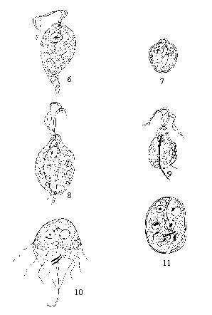
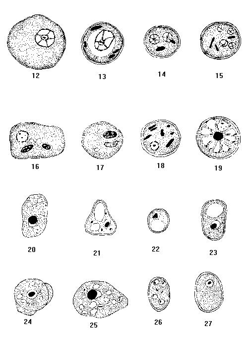
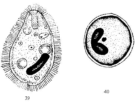
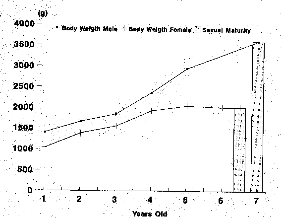
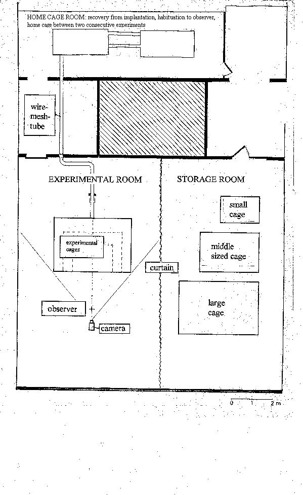
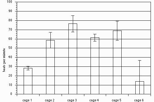