(These cases are from Dr. Kessimian’s teaching archives back during the 1980’s -1990’s. They are presented here for both educational and historical purposes.)
Case 1: Metastatic adenocarcinoma (Liver biopsy)
Case 2: Metastatic esophageal adenocarcinoma (Mediastinal lymph node)
Case 3: Goblet cells of colon
Case 4: Metastatic atypical carcinoid (Cervical lymph node)
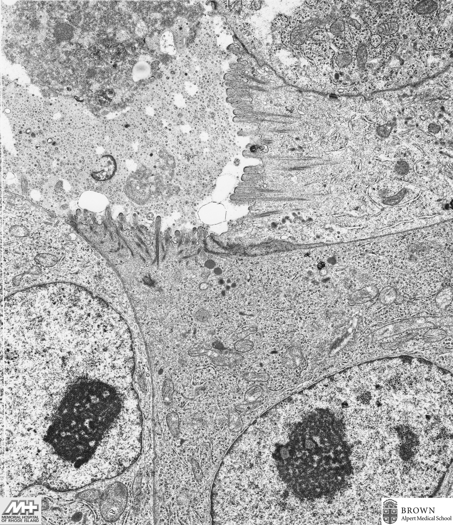
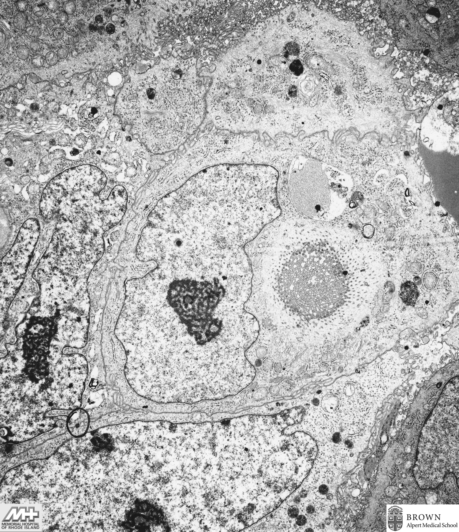
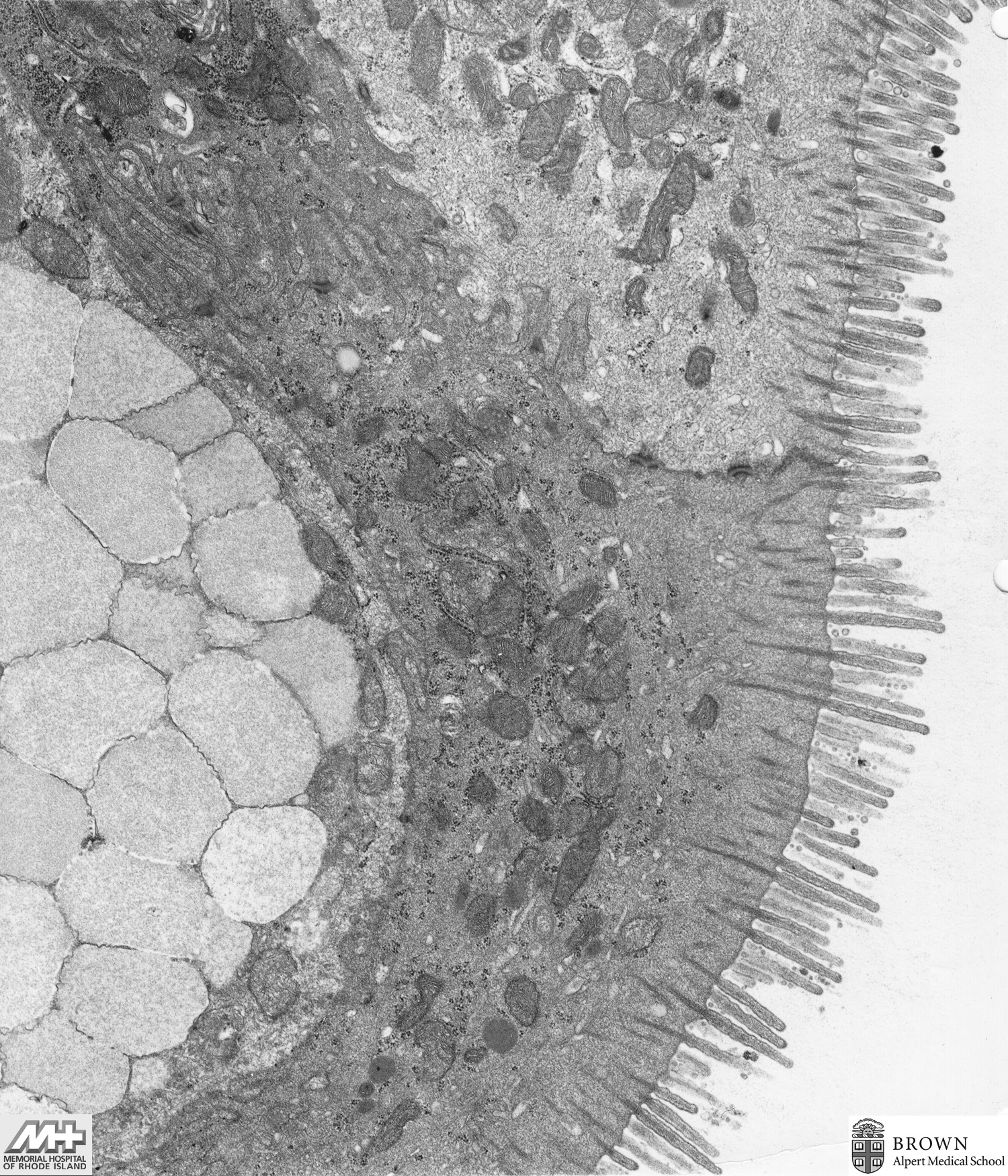
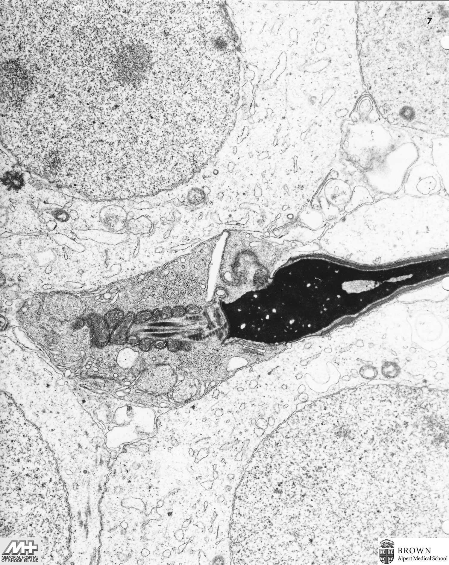
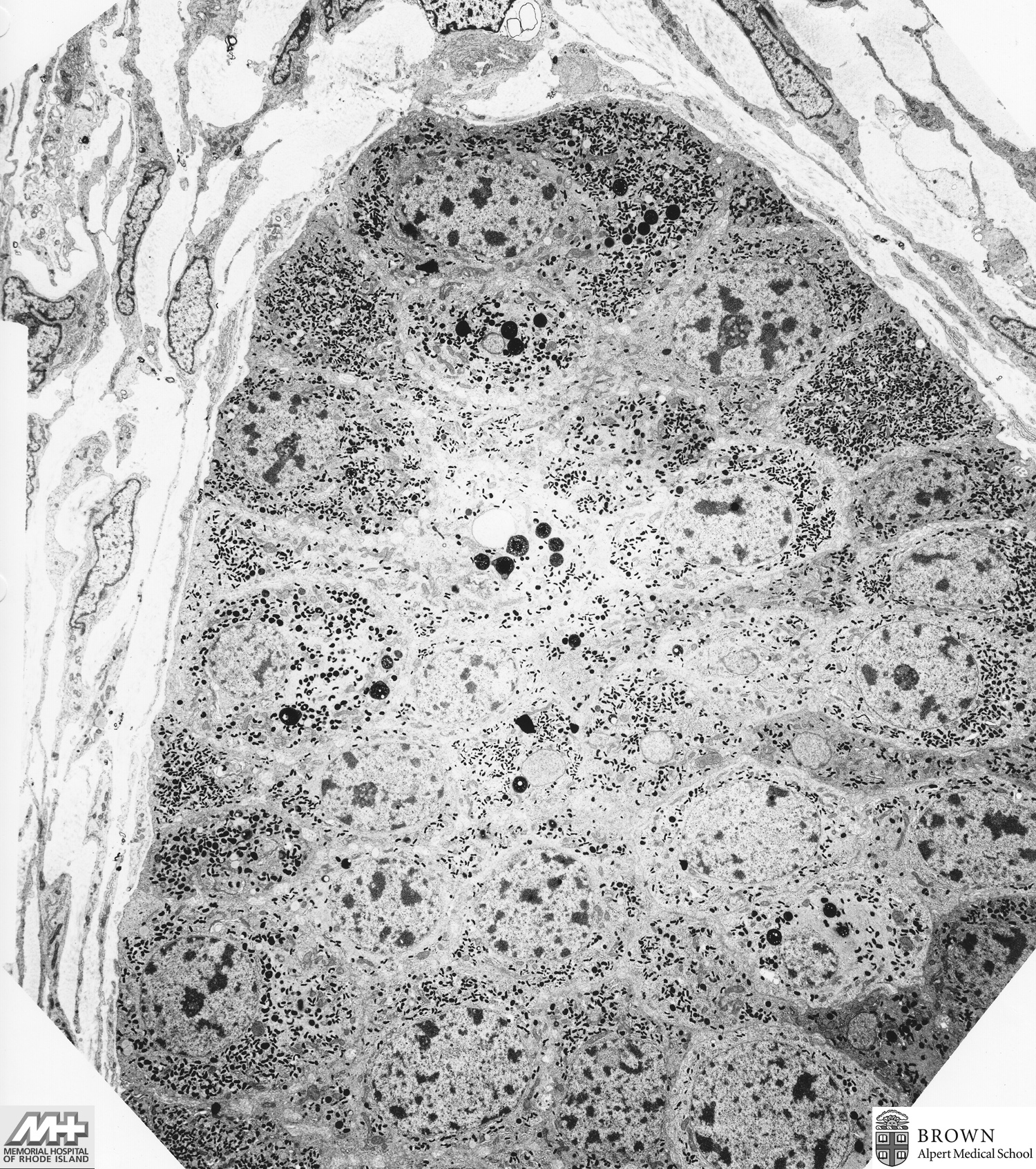
Case 5: Normal sperm (Testis biopsy)
Case 6: Normal pituitary
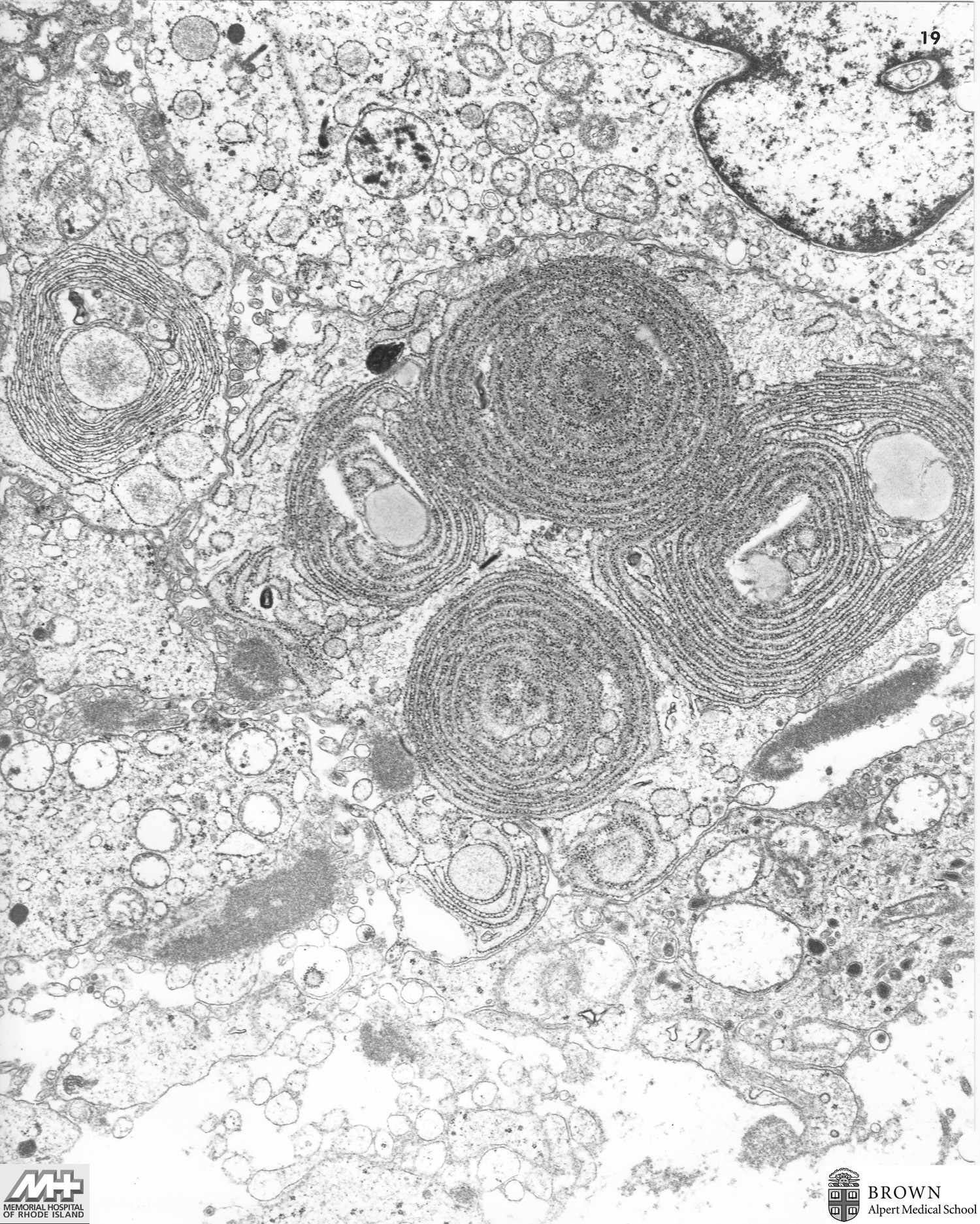
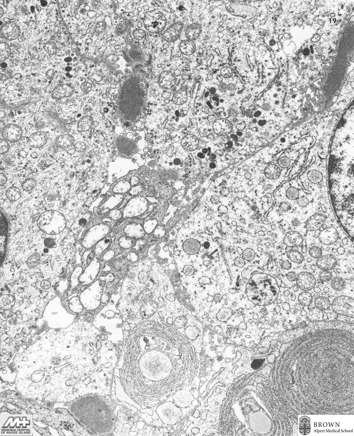
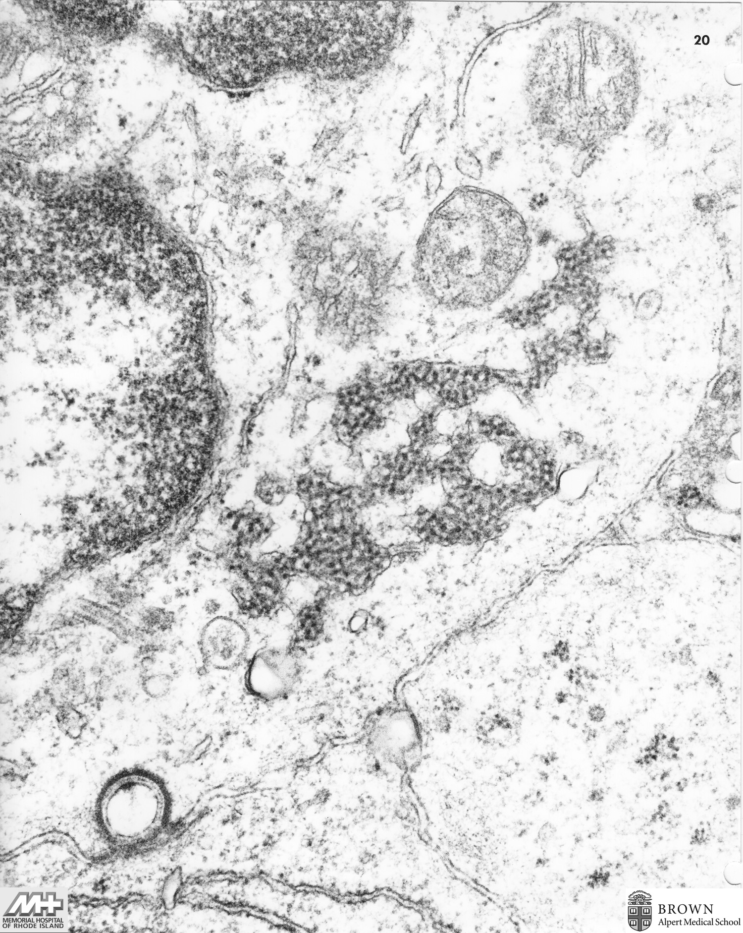
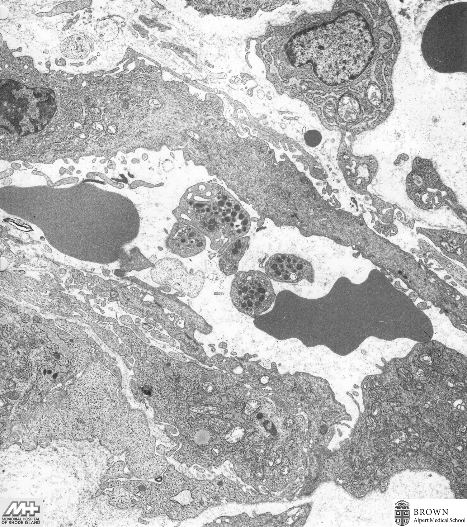
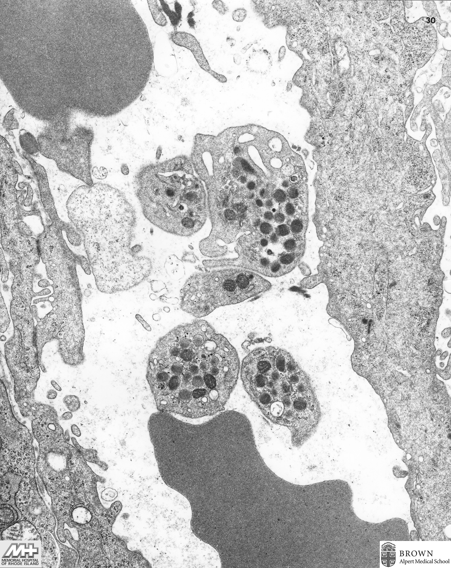
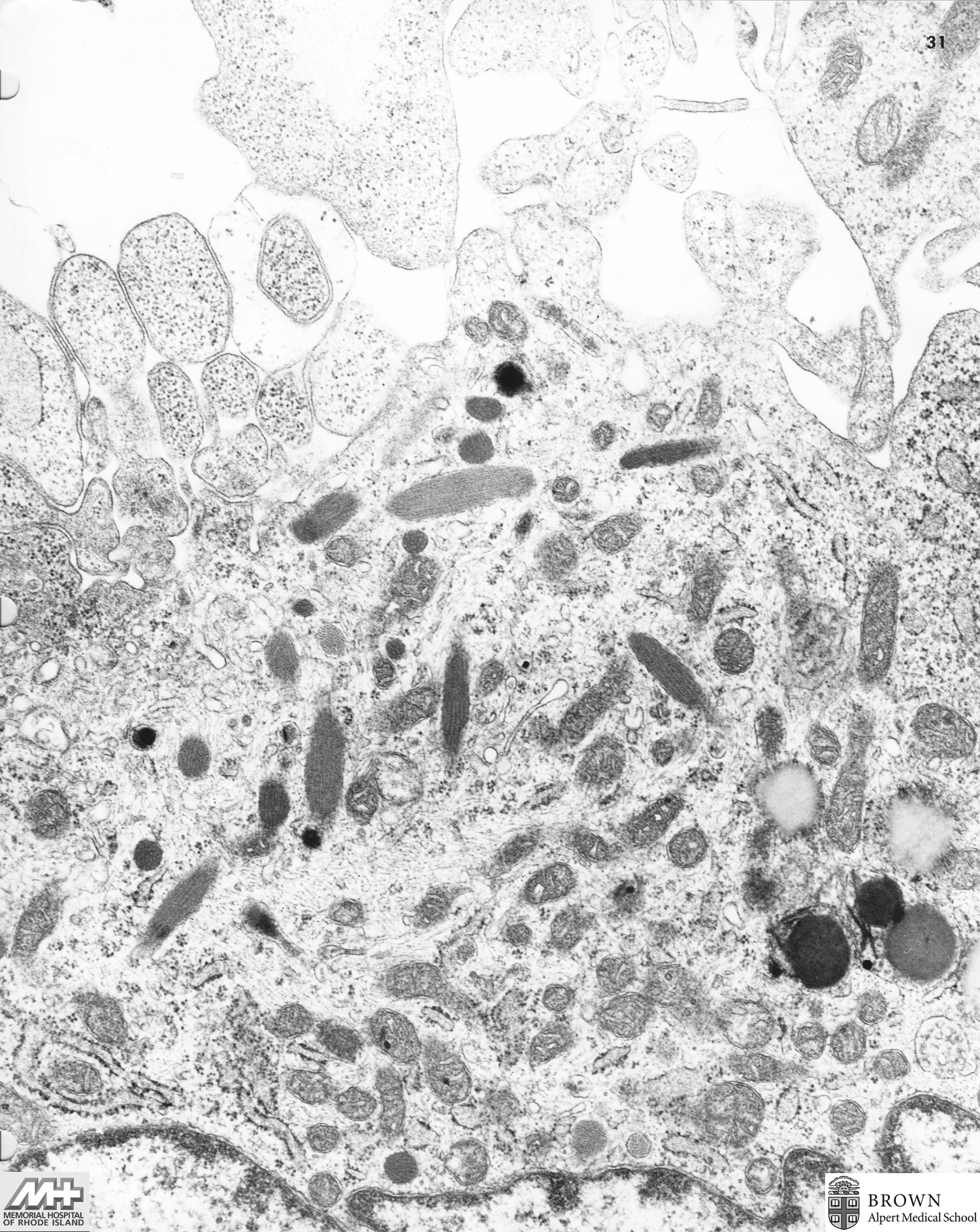
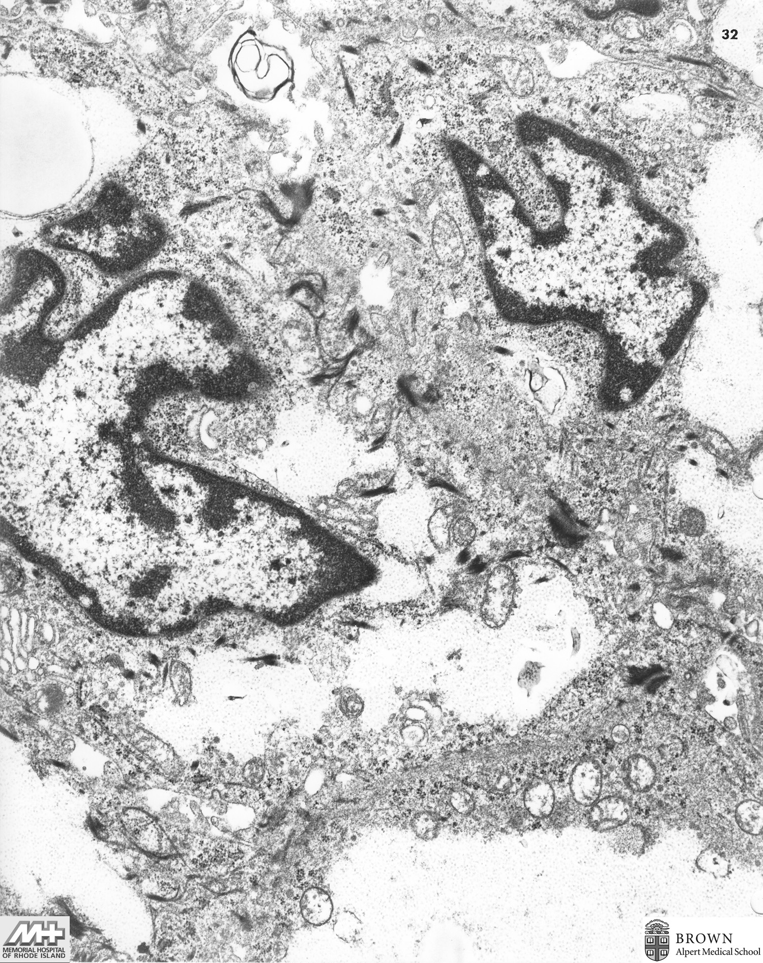
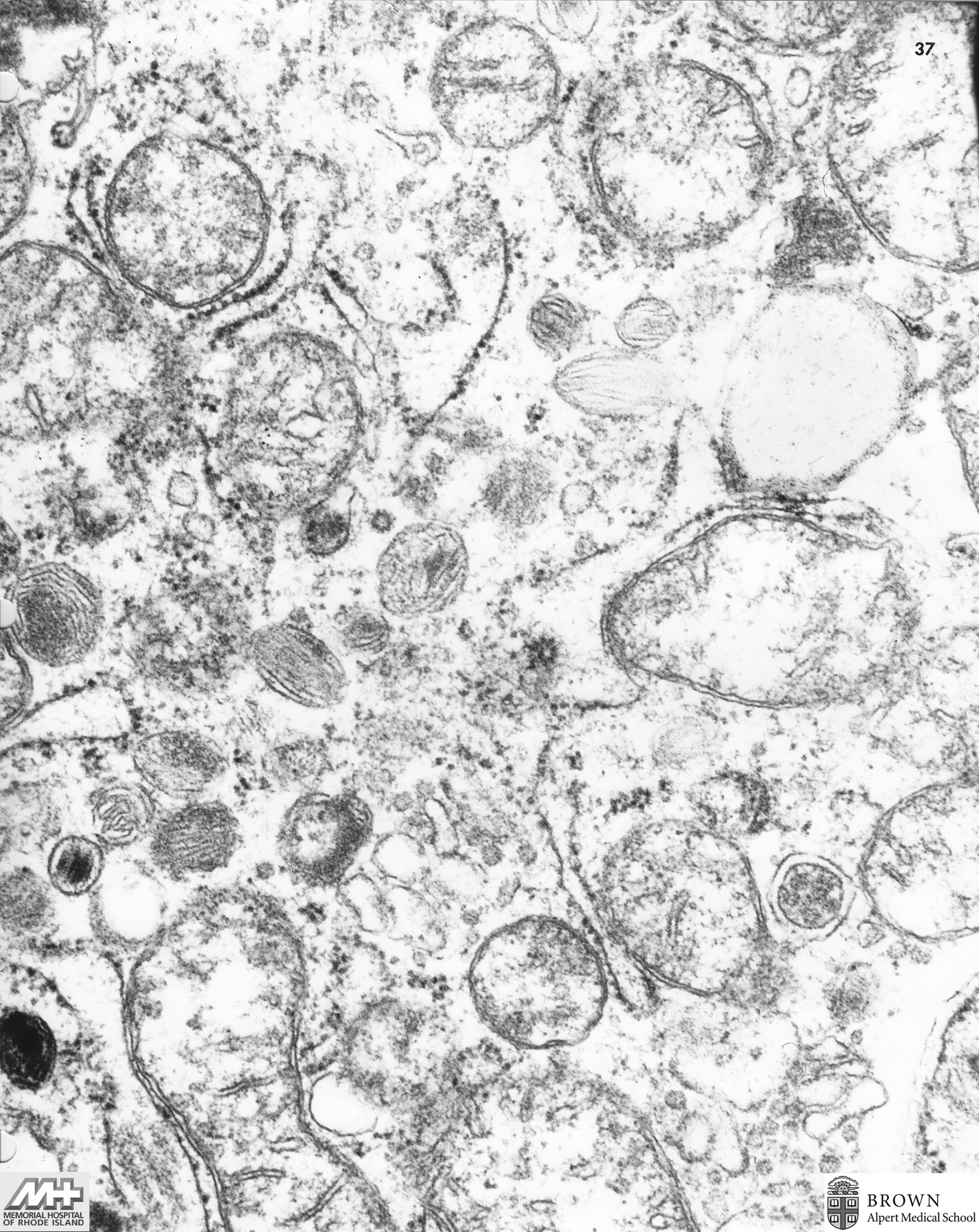
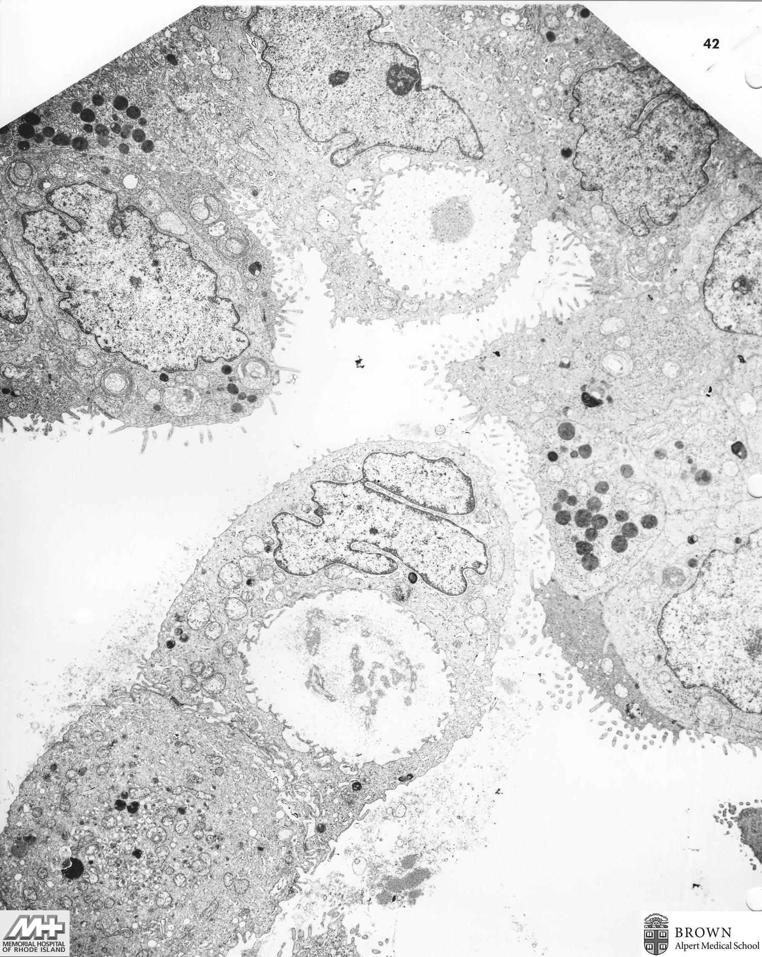
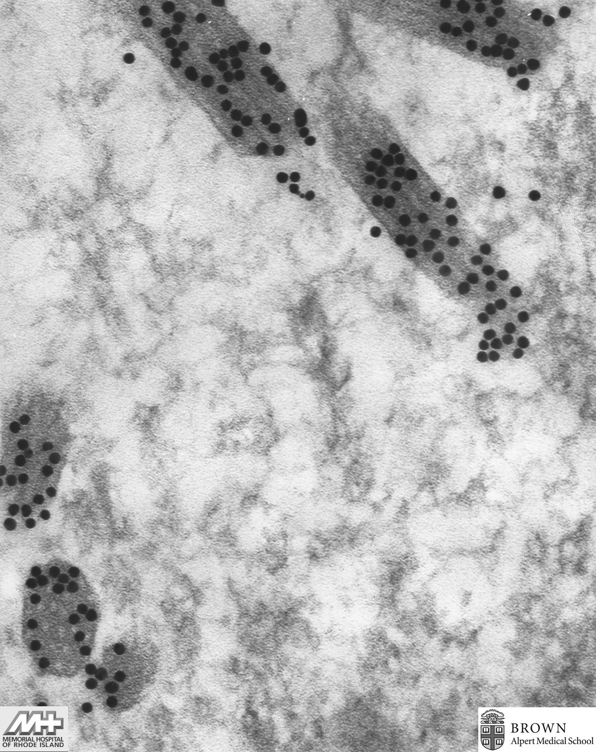
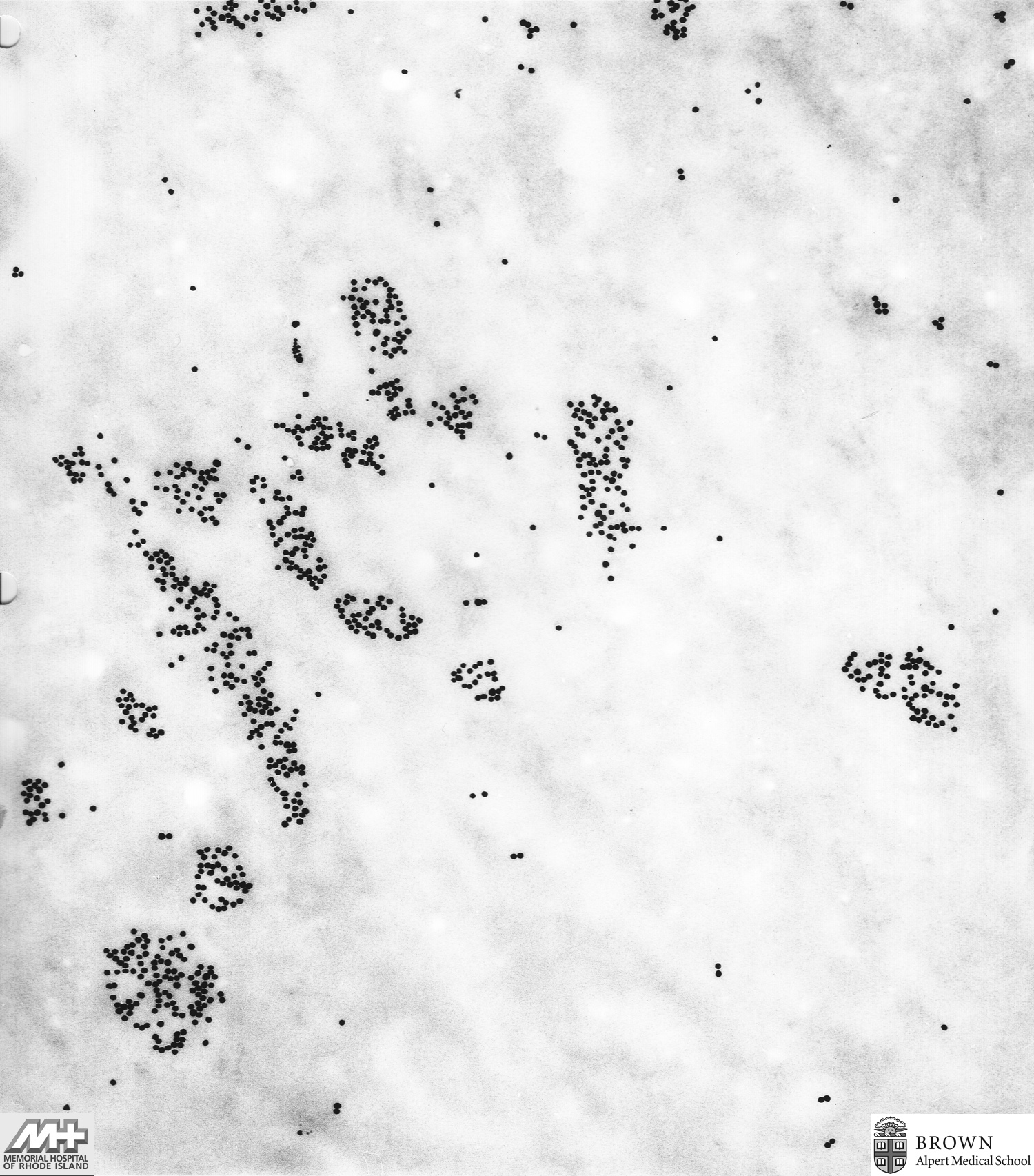
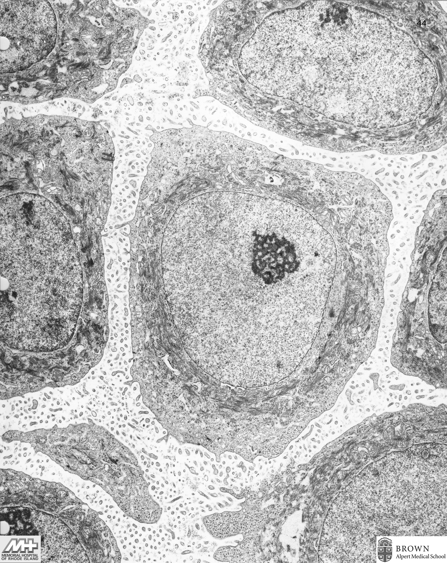
Case 7: Toxoplasmosis (Brain)
Case 8: Hepatocellular carcinoma (Liver)
Case 9: Metastatic adenocarcinoma (Liver)
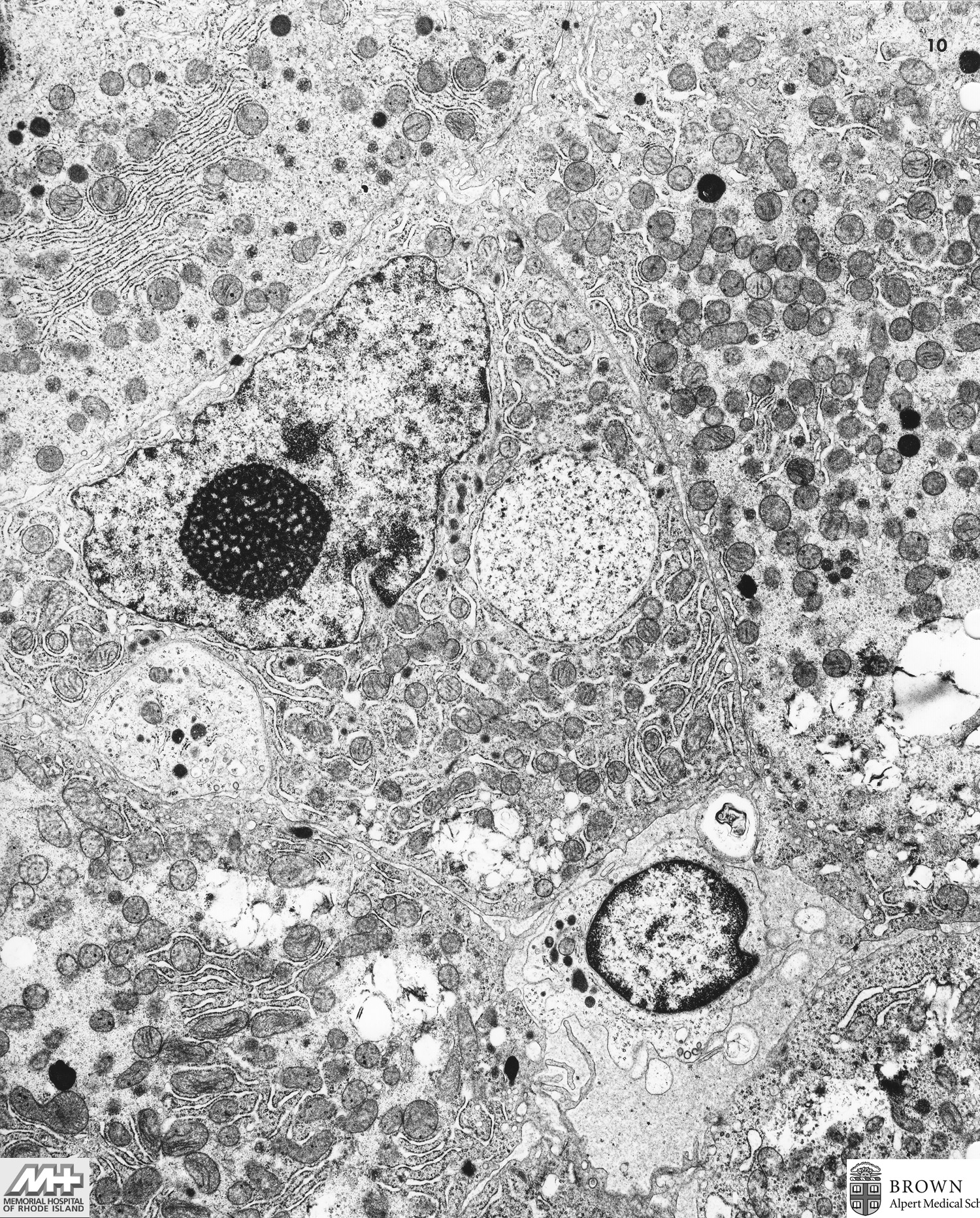
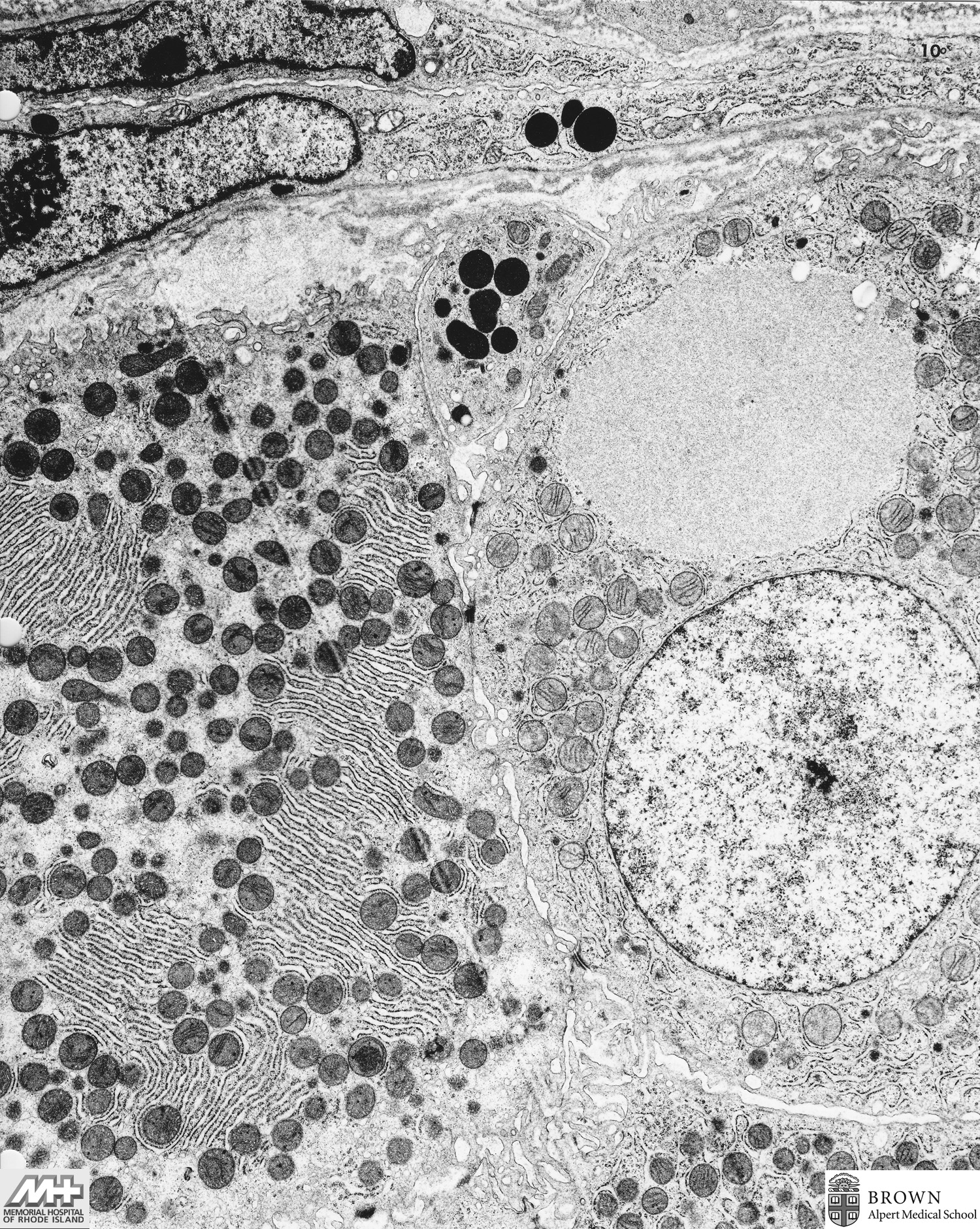
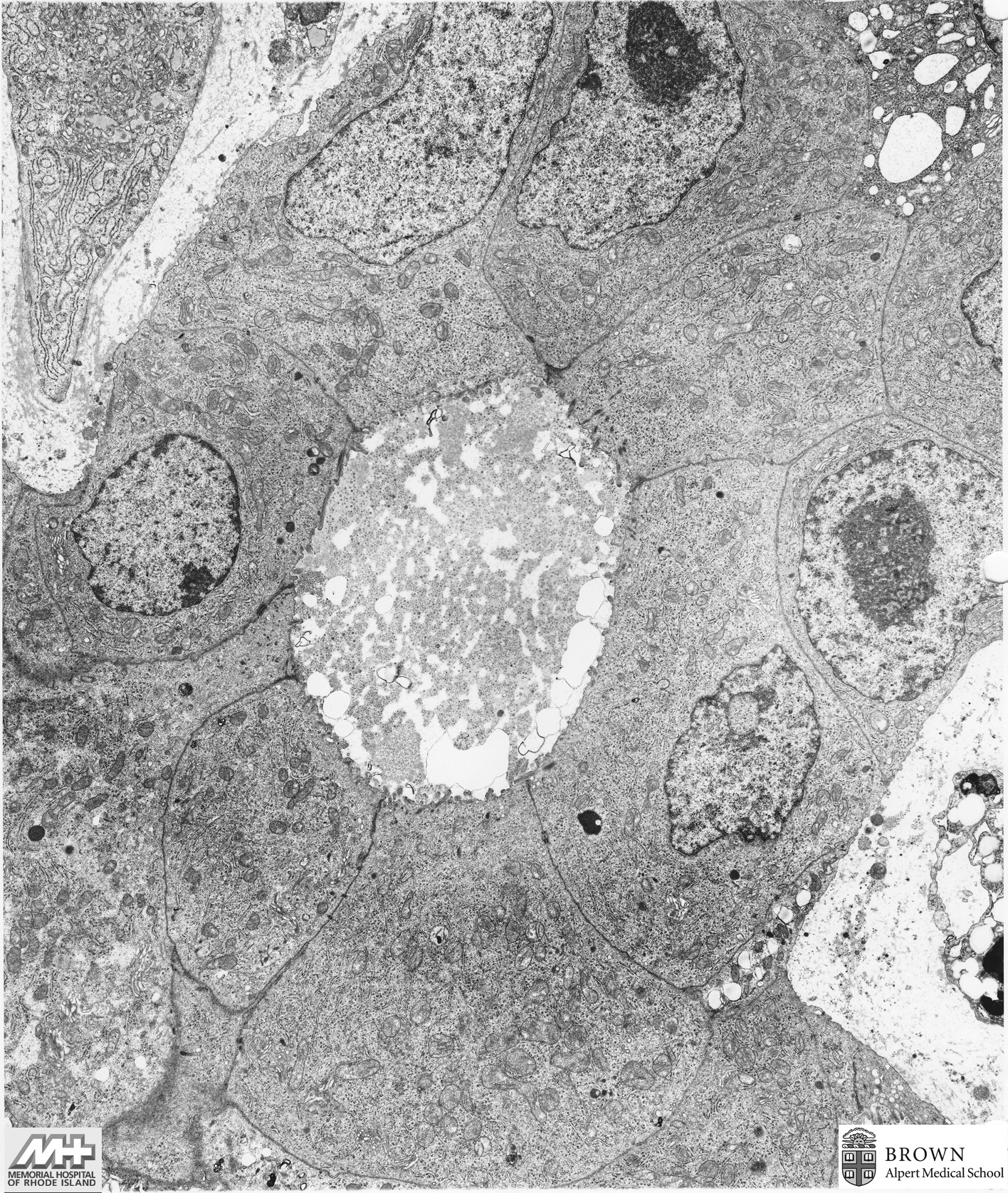
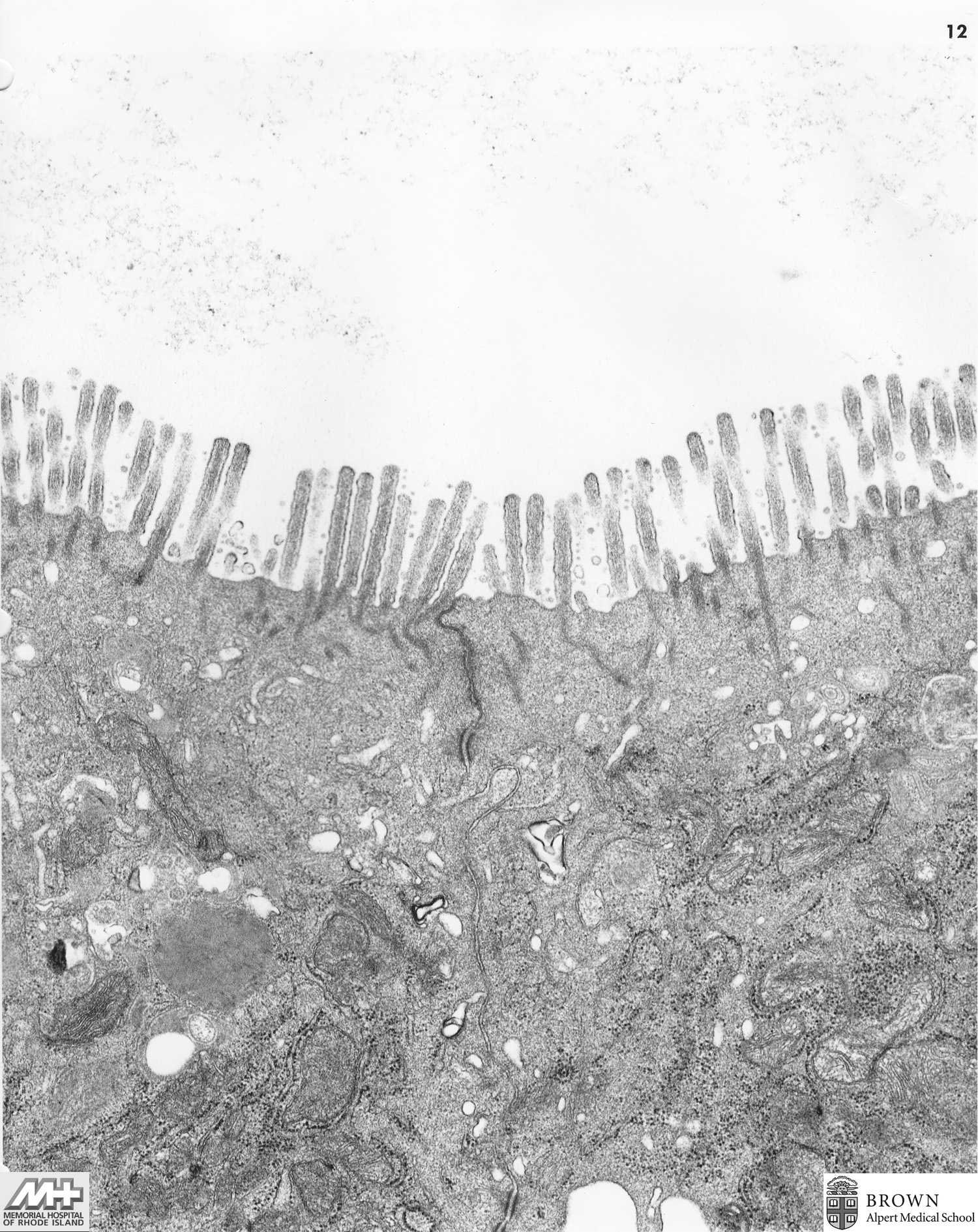
Case 10: Adenocarcinoma in tubulovillous adenoma (Colonic polyposis)
Case 11: Paget's disease (Anus)
Case 12: Bronchioloalveolar Carcinoma (Lung)
Case 13: Carcinoid (Appendix)
Case 14: Schwannoma (Luse bodies)
Case 15: Clear cell adenocarcinoma (Vagina)
Case 16: Oat cell carcinoma (Cervical lymph node)
Case 17: Metastatic melanoma (Axilla)
Image 1 (4,300x) Image 2 (10,500x) Image 3 (35,000x) Image 4 (17,500x) Image 5 (17,500x)
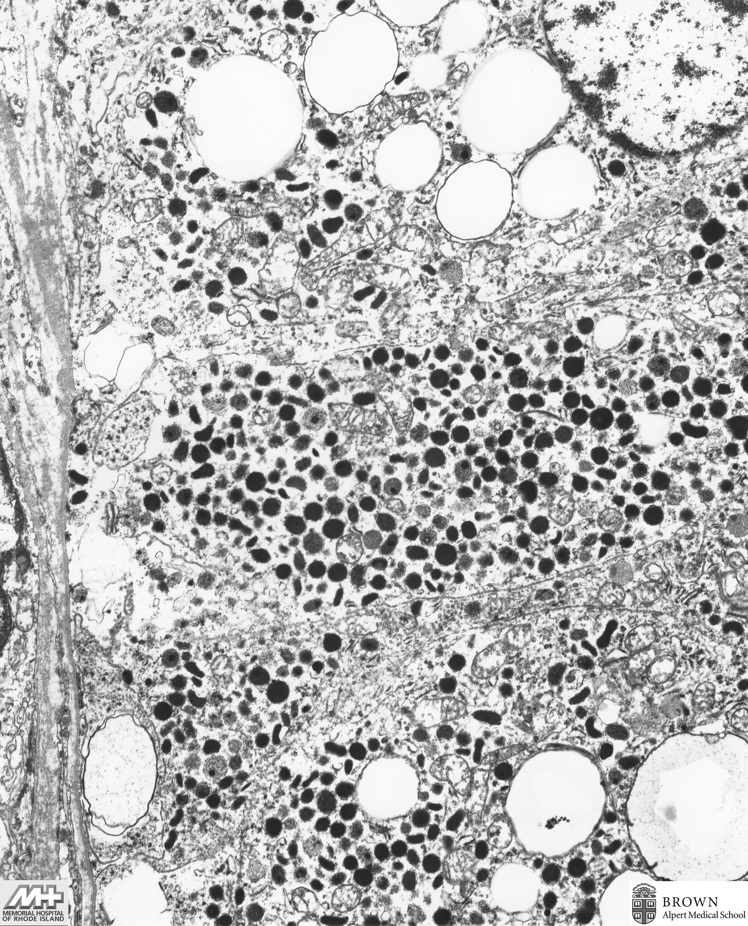
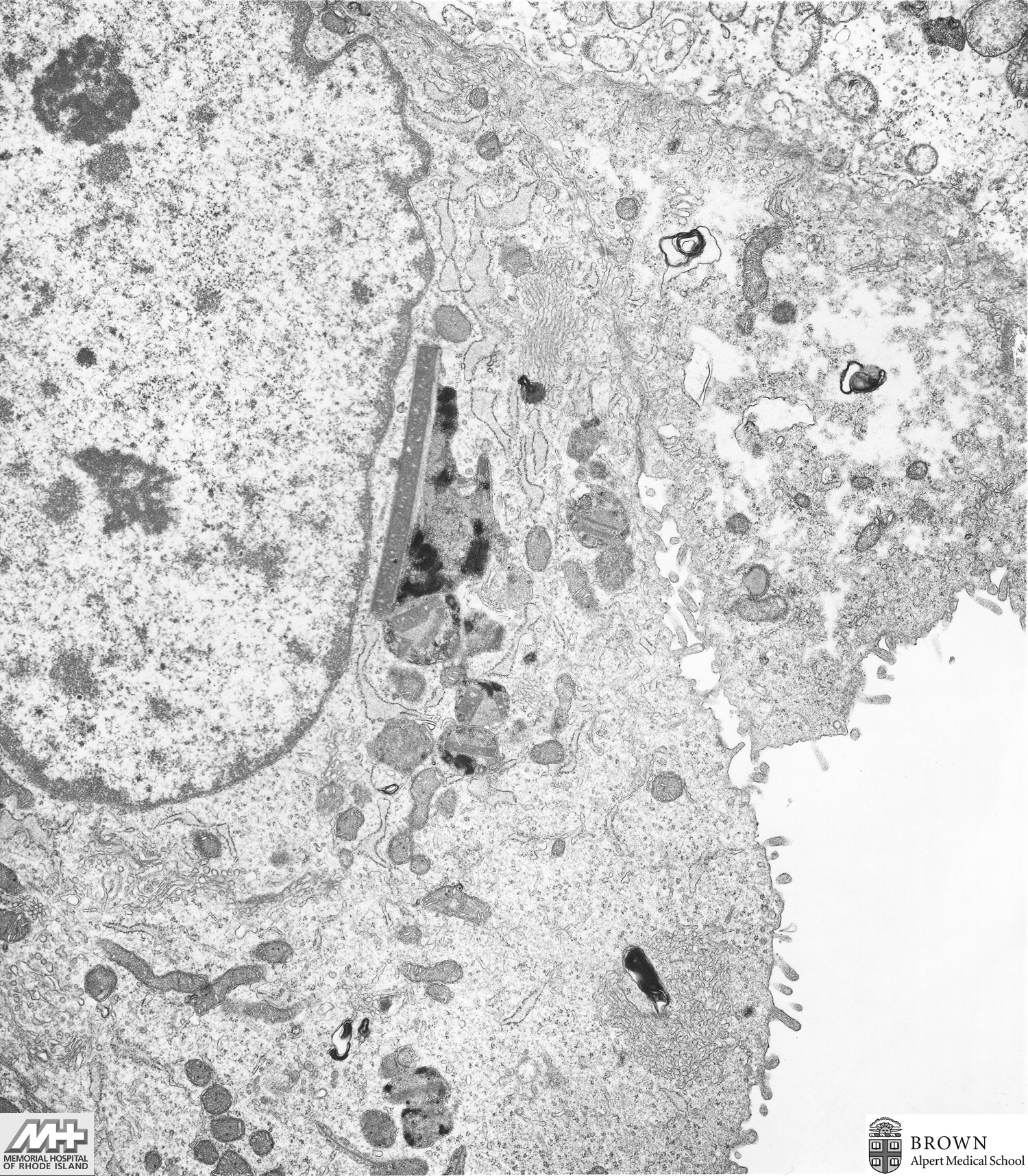
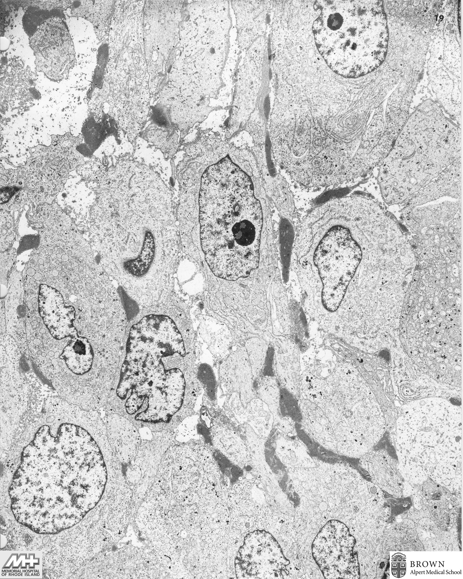
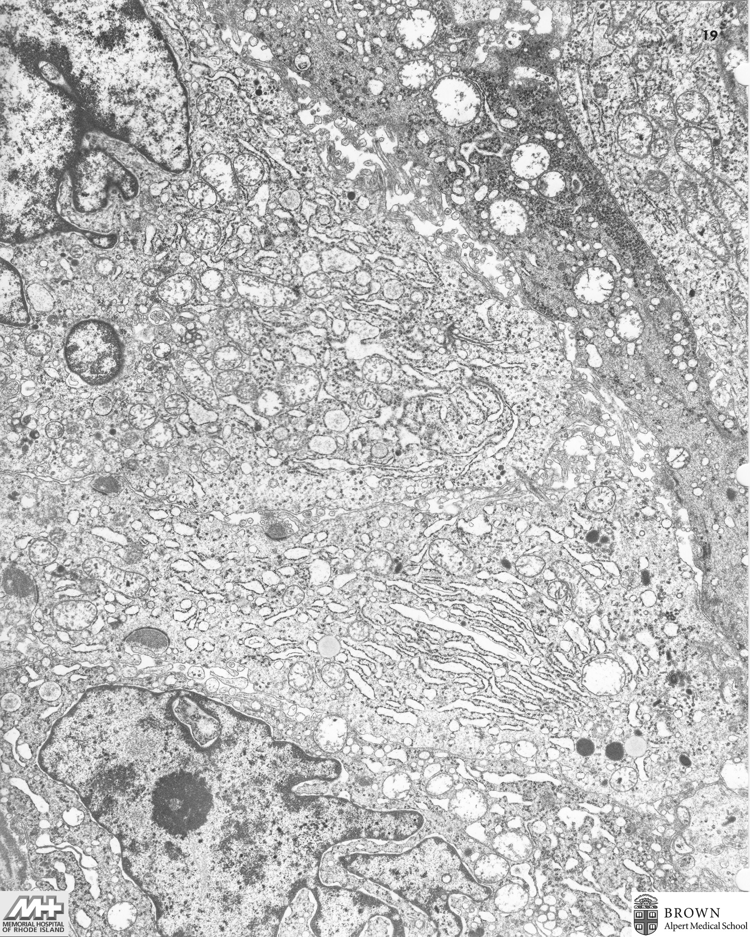
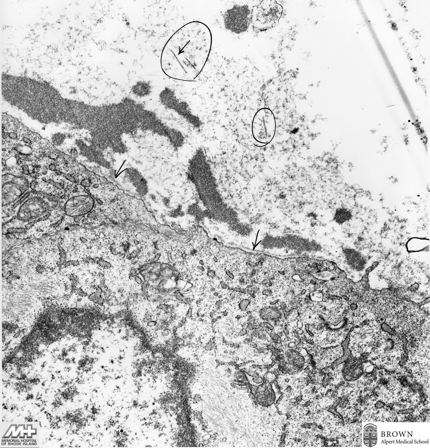
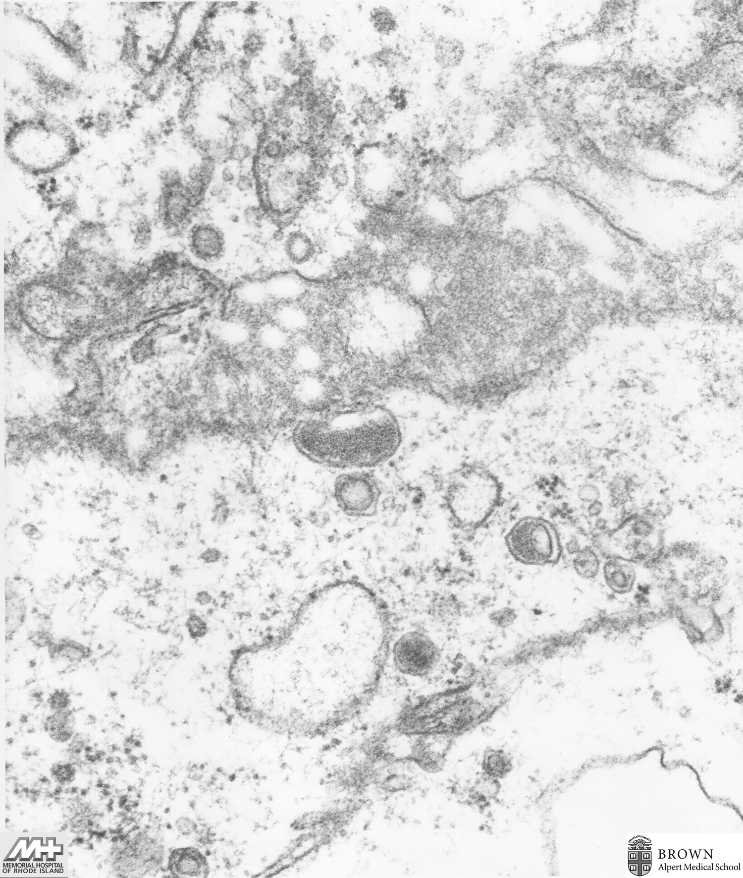
Case 18: Tubuloreticular structures associated with AIDS (HIV-positive male, intravenous drug abuser)
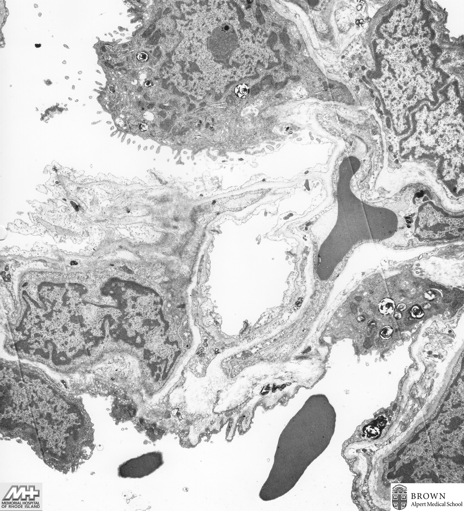
Case 19: Adenocarcinoma (Lung)
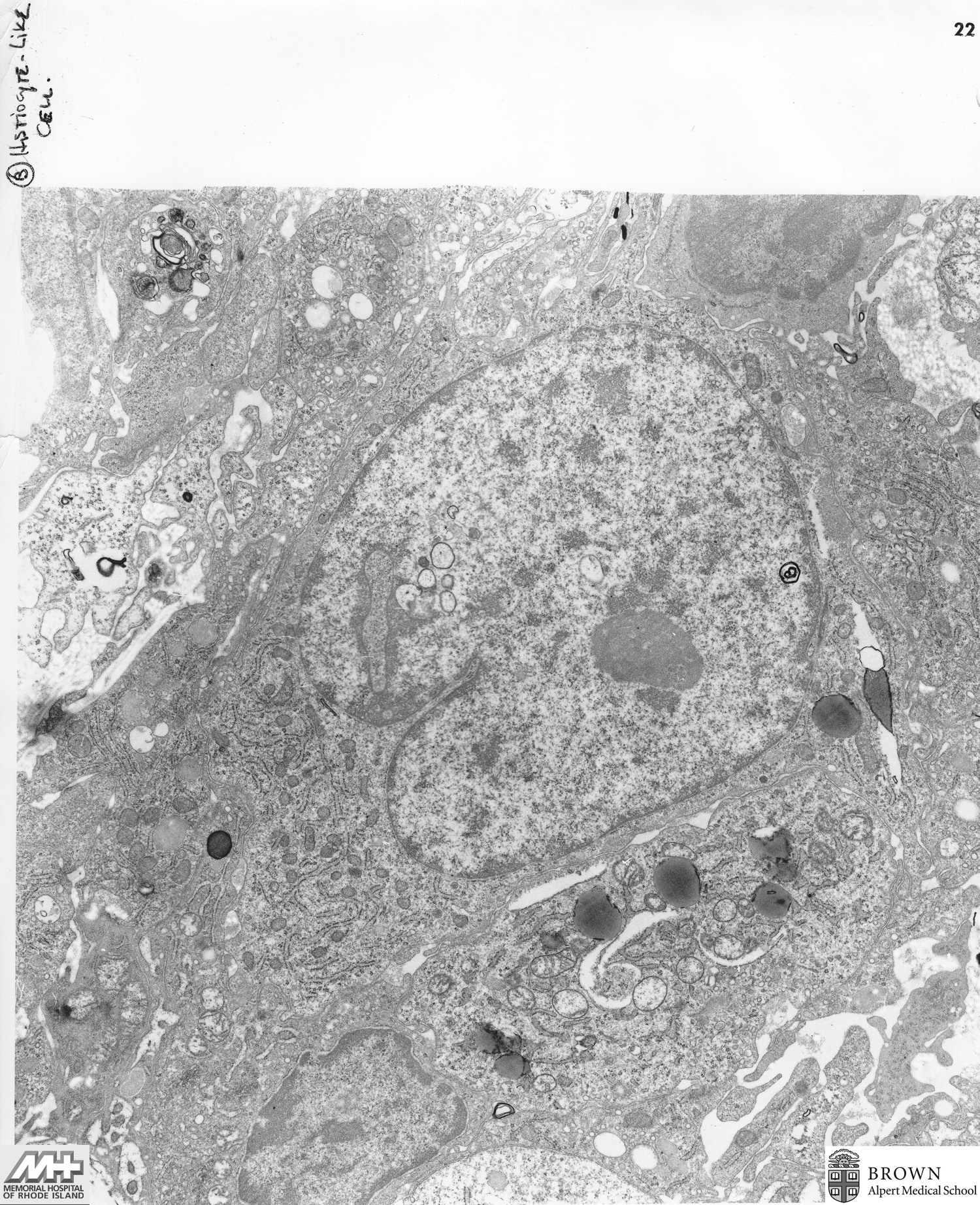
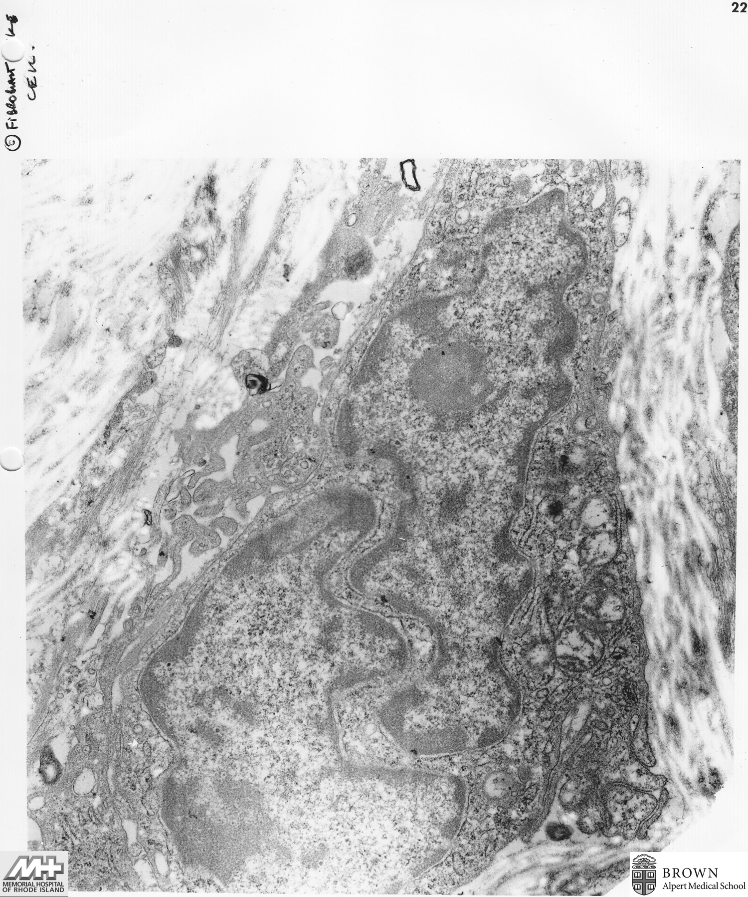
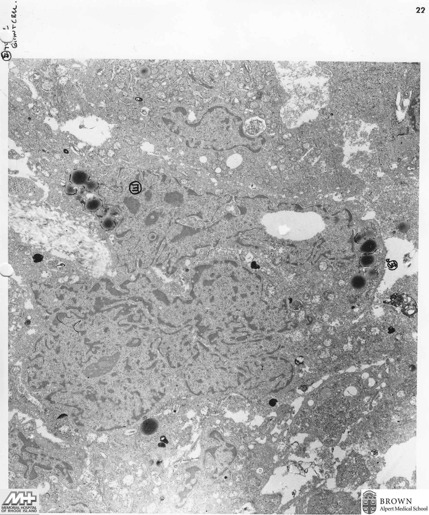
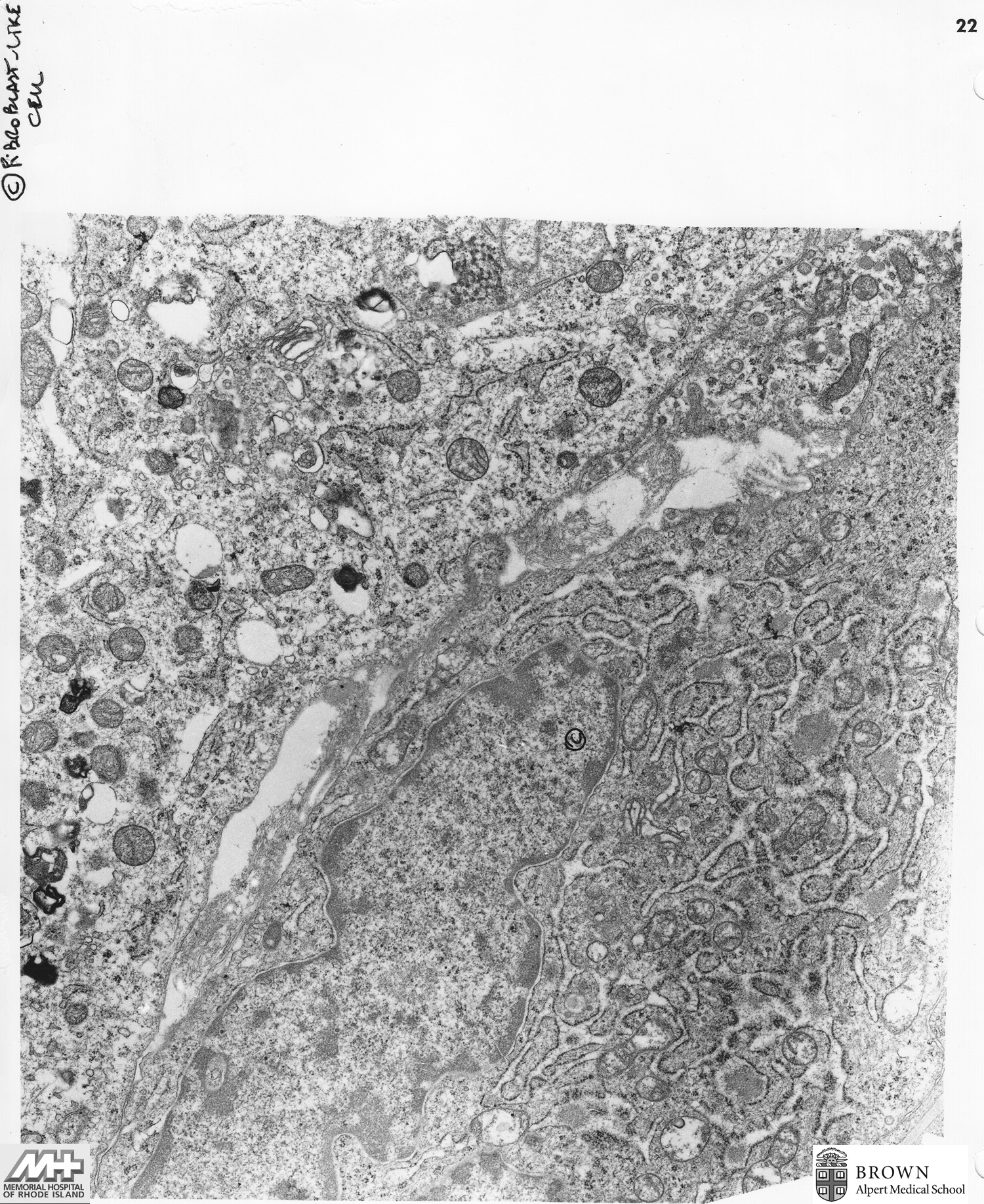
Case 20: Malignant Fibrohistiocytoma (Leg)
Image 1 (8,400x) Image 2 (15,000x) Image 3 (30,000x) Image 4 (19,600x) Image 5 (19,600x)
Case 21: Mucoepidermoid carcinoma (Submandibular gland)
Case 22: Malignant mesothelioma (Pleural biopsy)
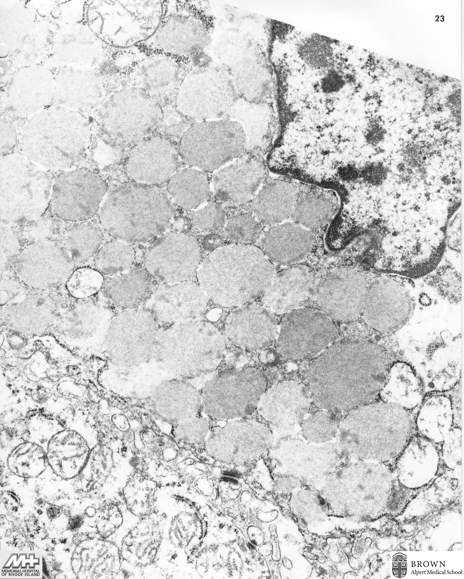
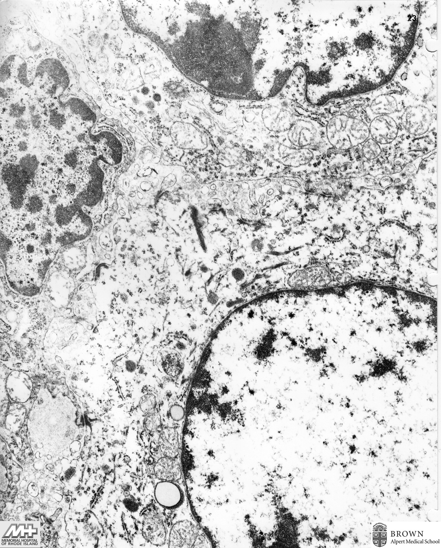
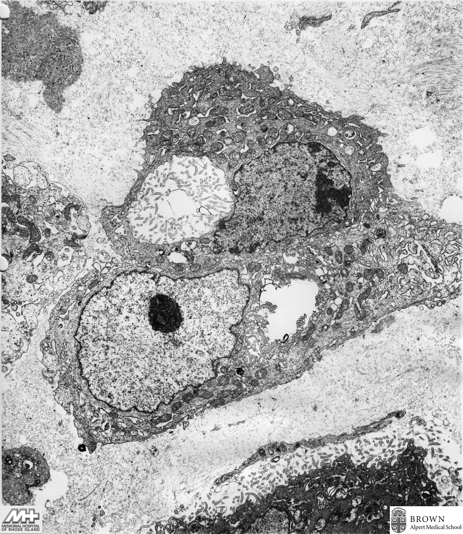
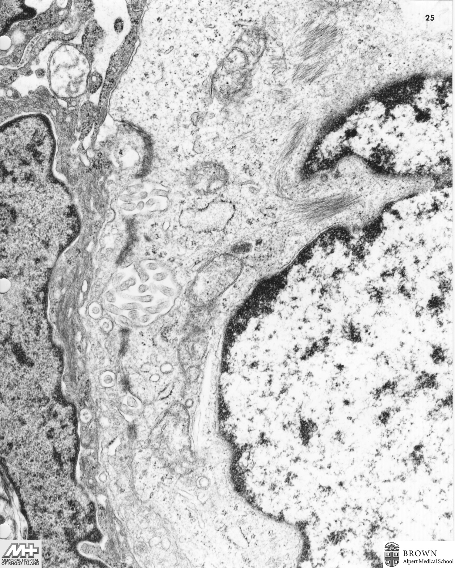
Case 23: Malignant mesothelioma (Pleural biopsy)
Case 24: Metastatic melanoma (Scapular region)
Case 25: Transtional cell carcinoma vs Mesothelioma (Anemone cell) (Peritoneum)
Case 26: Granular cell tumor (Finger)
Case 27: Small cell undifferentiated carcinoma (Mediastinal biopsy)
Case 28: Aggregate of platelets in vessel (Nodule of bowel)
Case 29: Melanotic neuroectodermal tumor of infancy (progonoma) (Maxilla)
Case 30: Intradermal nevus (Nose)
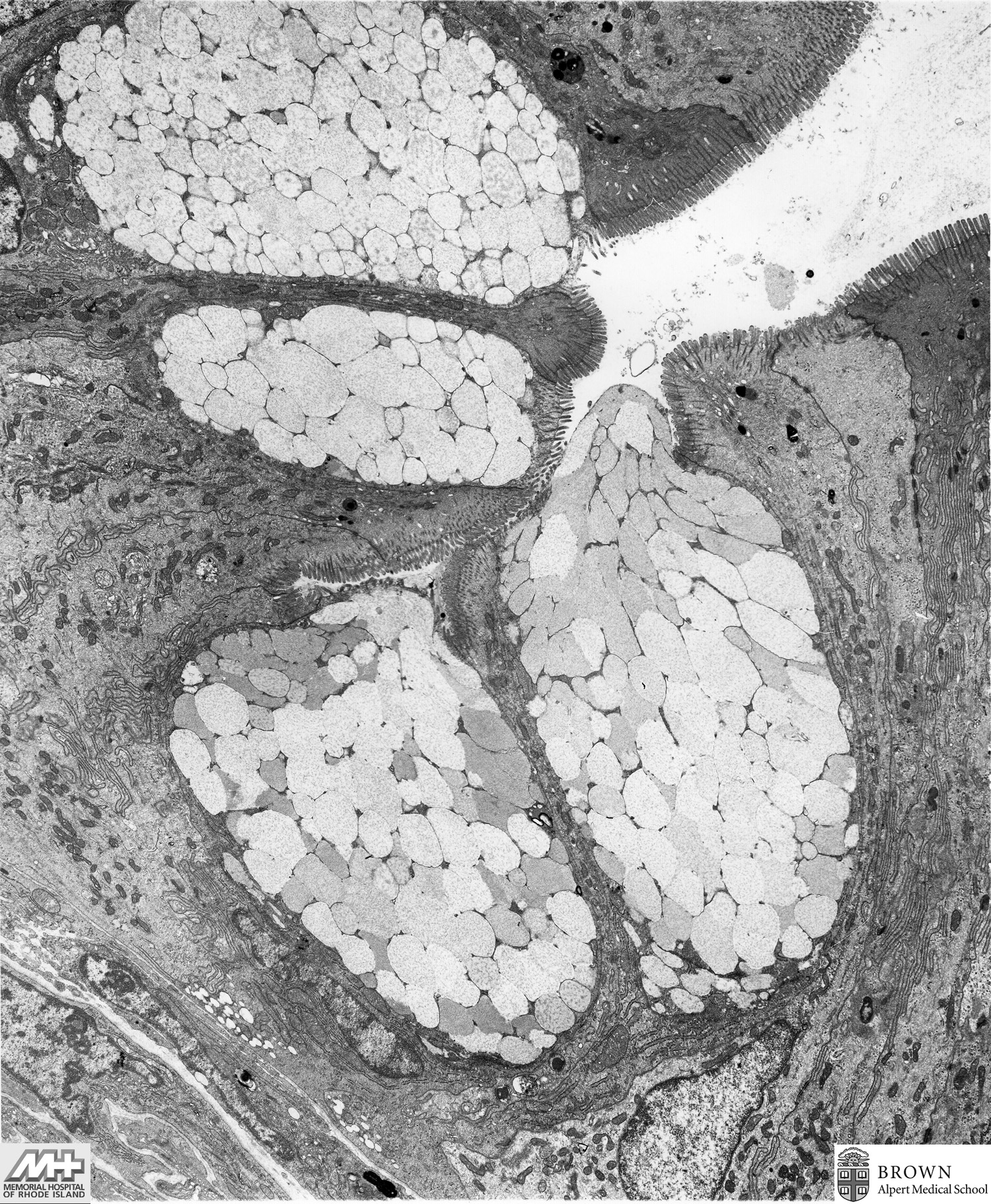
Case 31: Goblet cells in a tubular adenoma (Colonic biopsy)
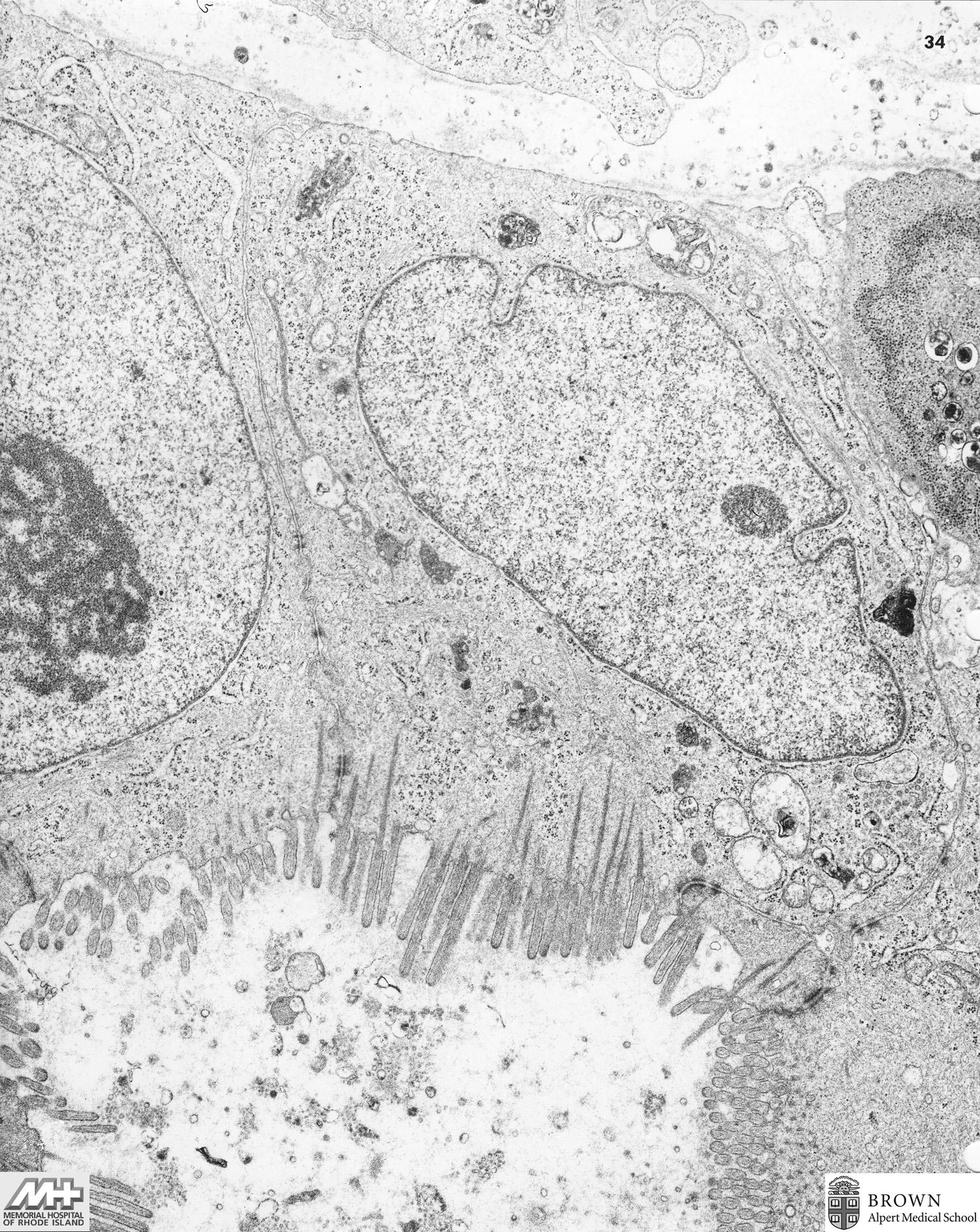
Case 32: Adenocarcinoma (Gall bladder)
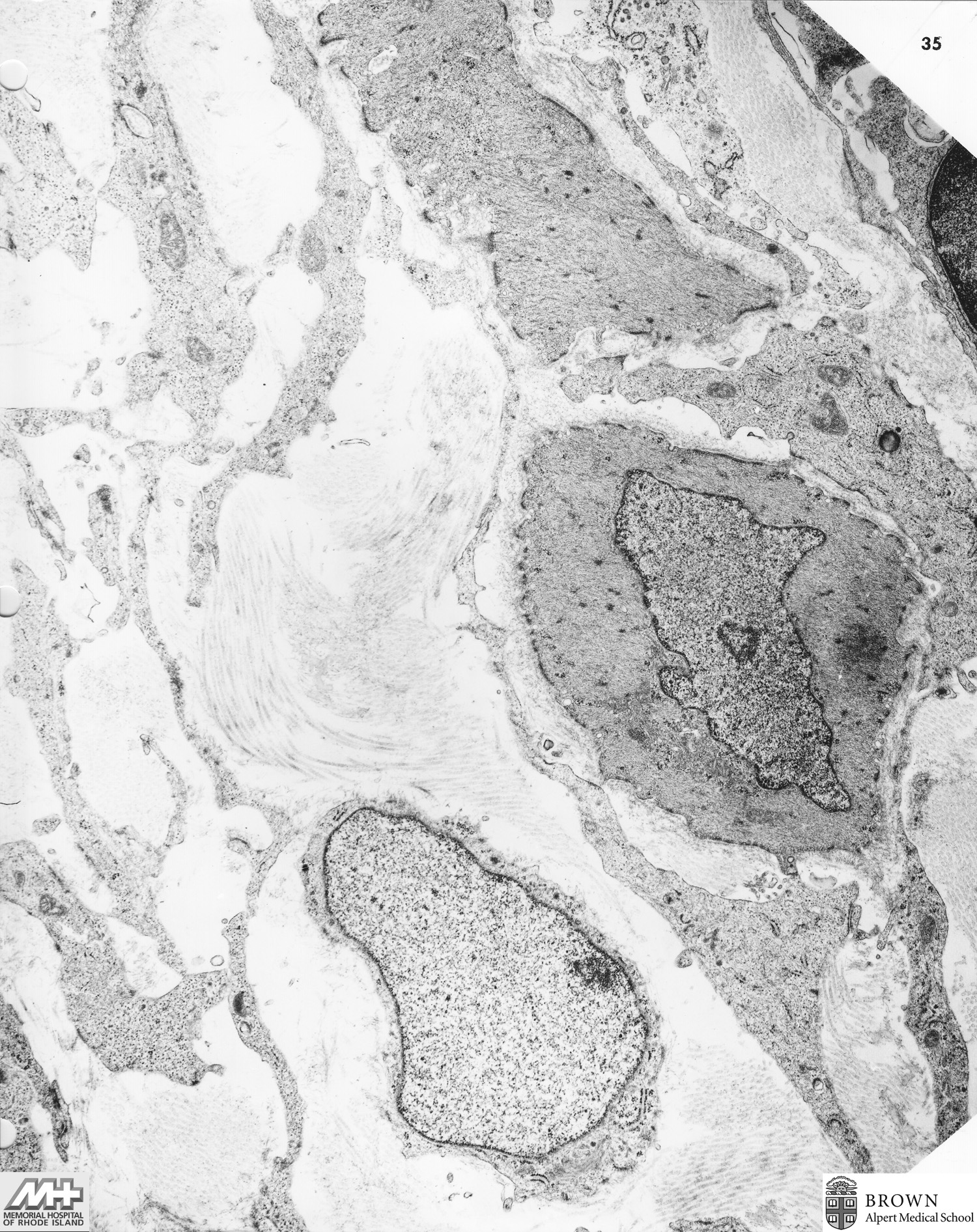
Case 33: Angiofibromatous hyperplasia with smooth muscle (Prostate)
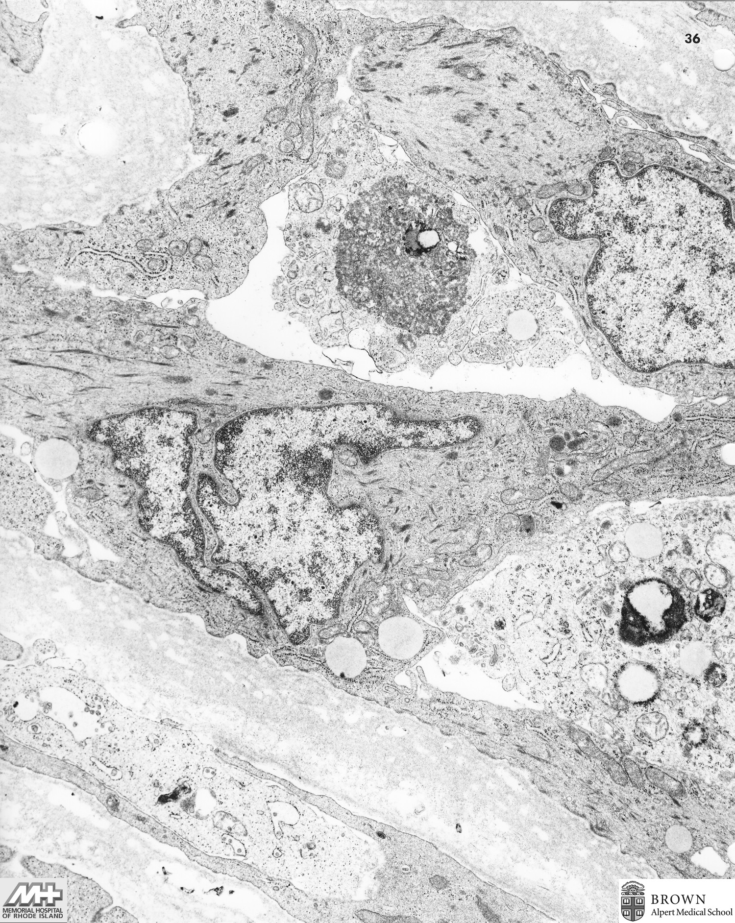
Case 34: Vessel wall with smooth muscle (Lung)
Case 35: Melanoma (skin)
Image 1 (10,500x) Image 2 (21,000x) Image 3 (70,000x) Image 4 (35,000x) Image 5 (70,000x)
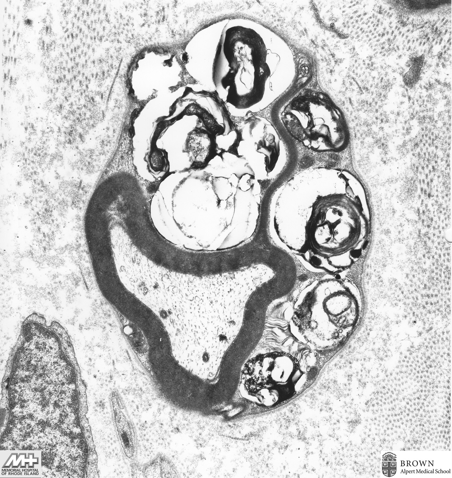
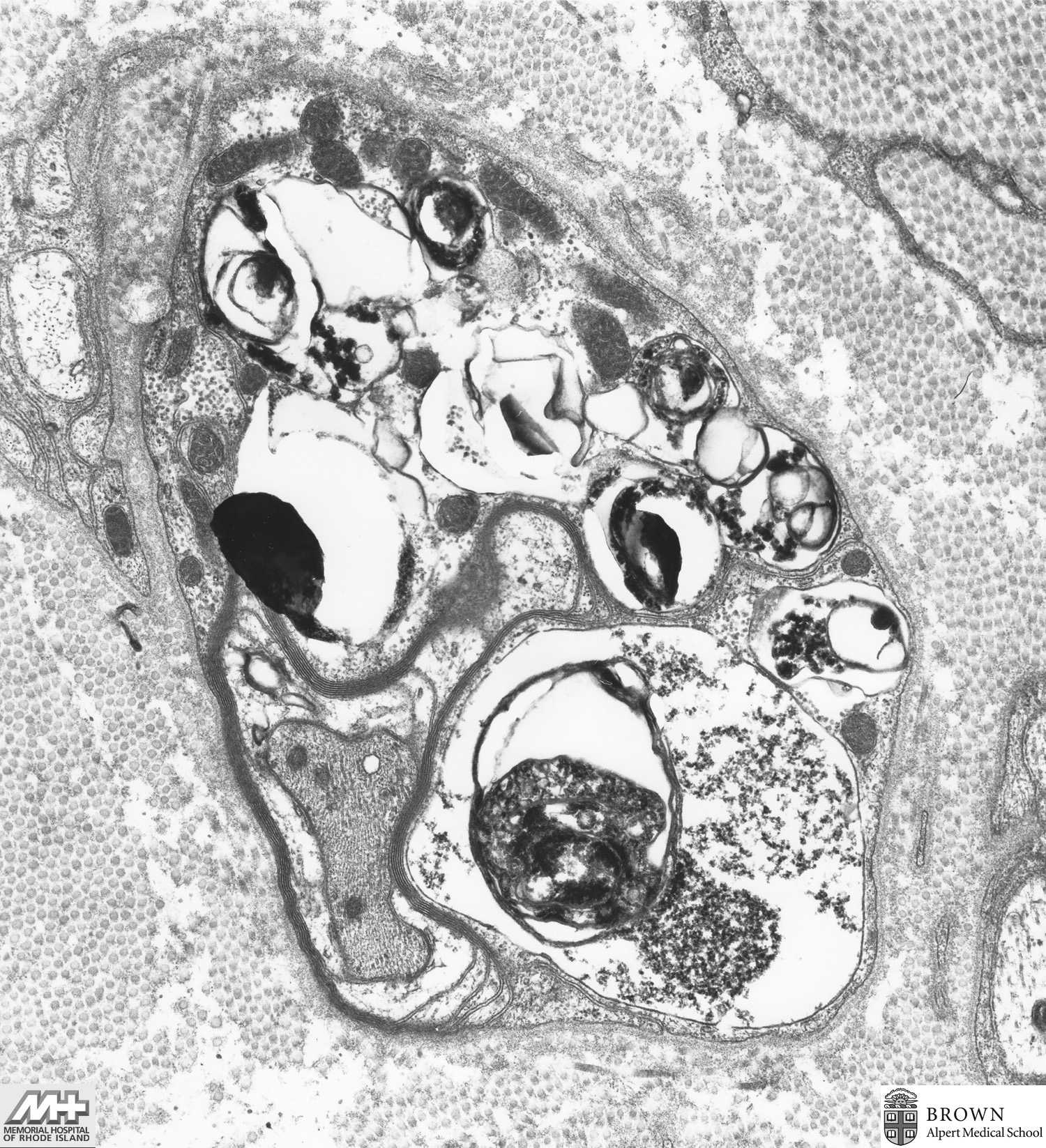
Case 36: Metachromatic leukodystrophy (Sural nerve)
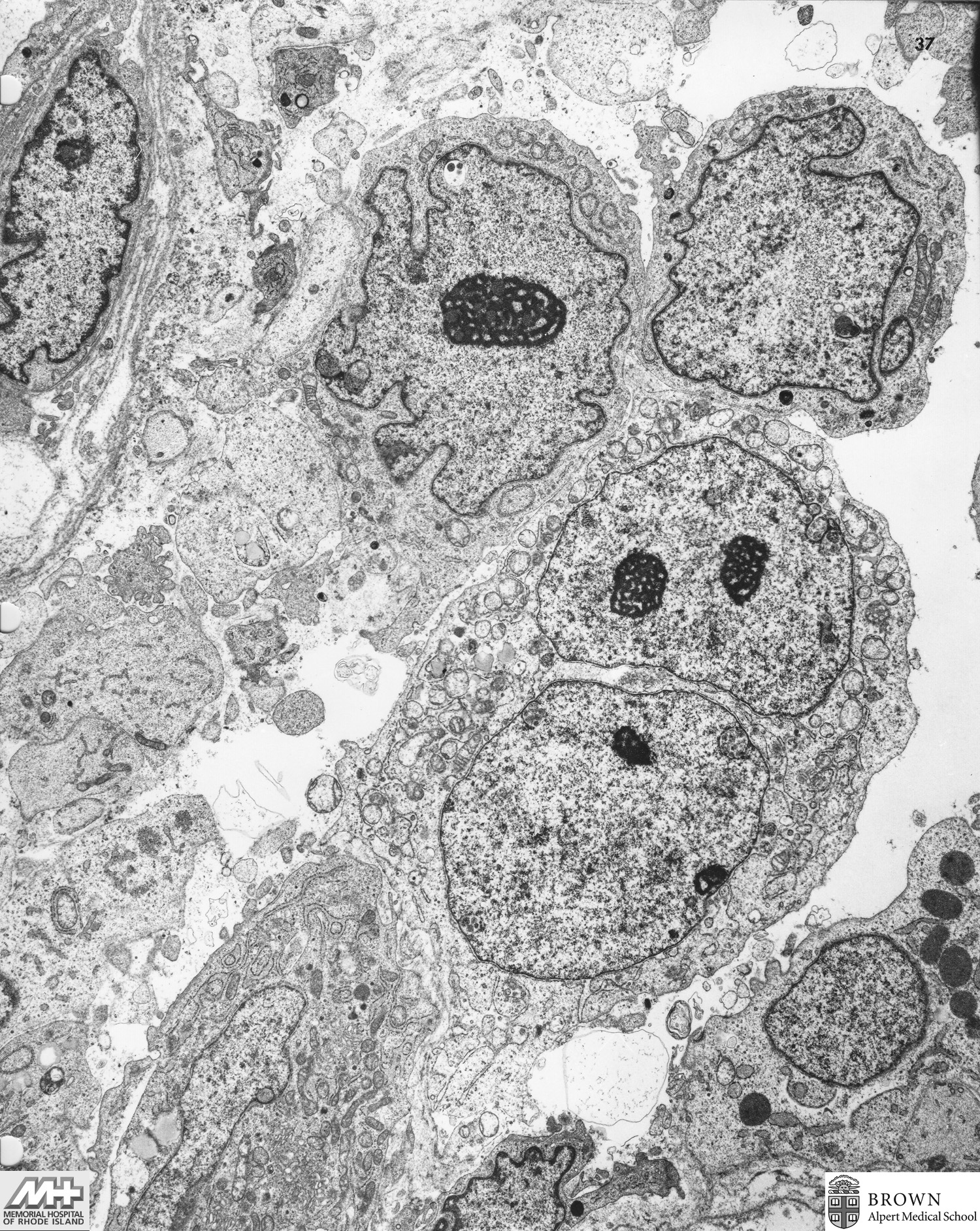
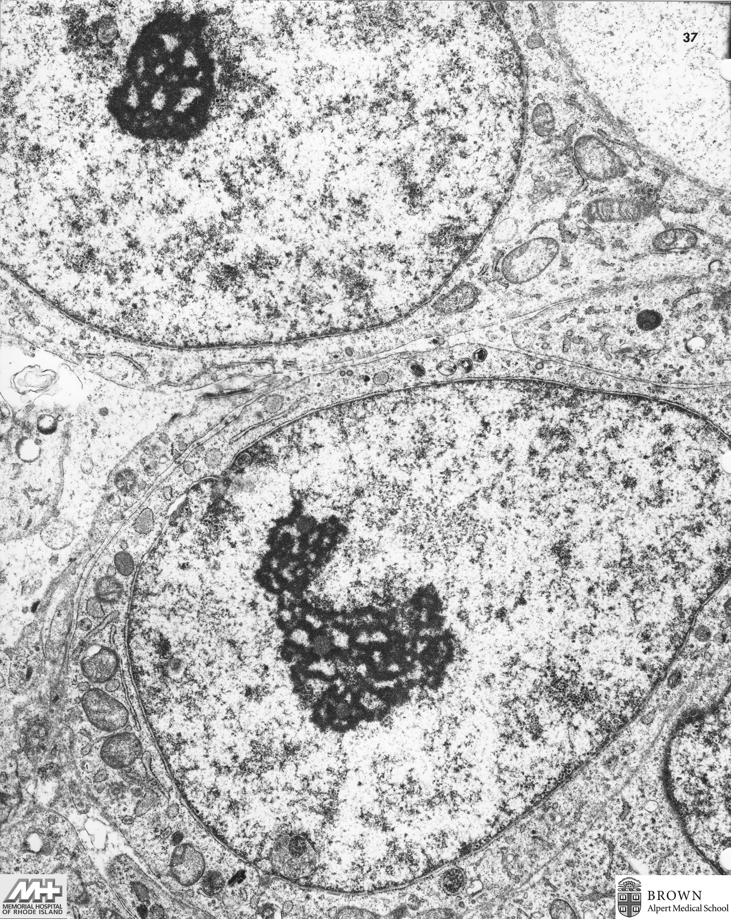
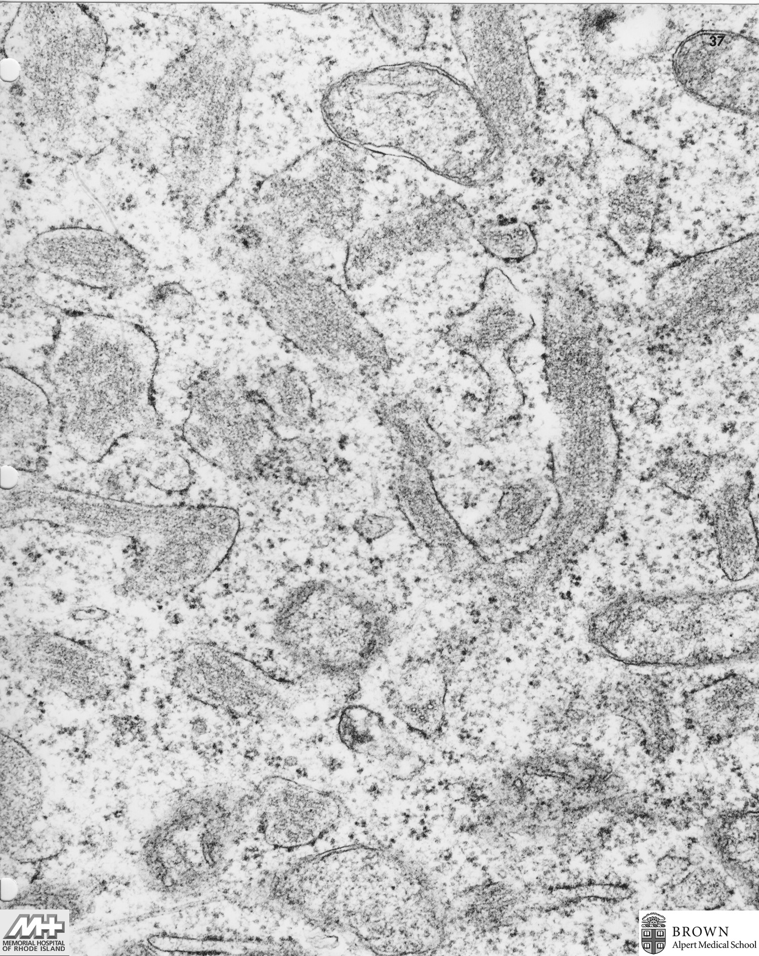
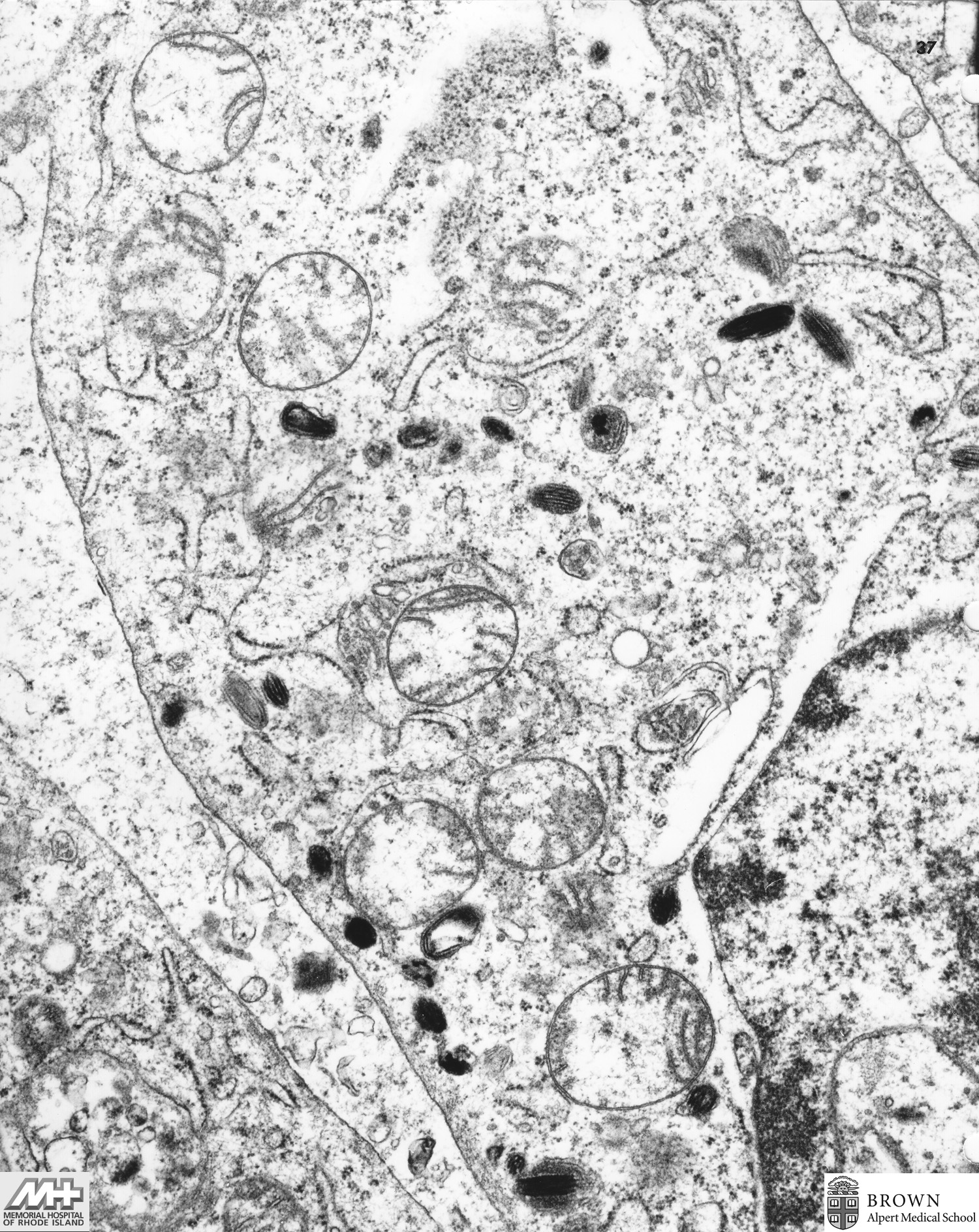
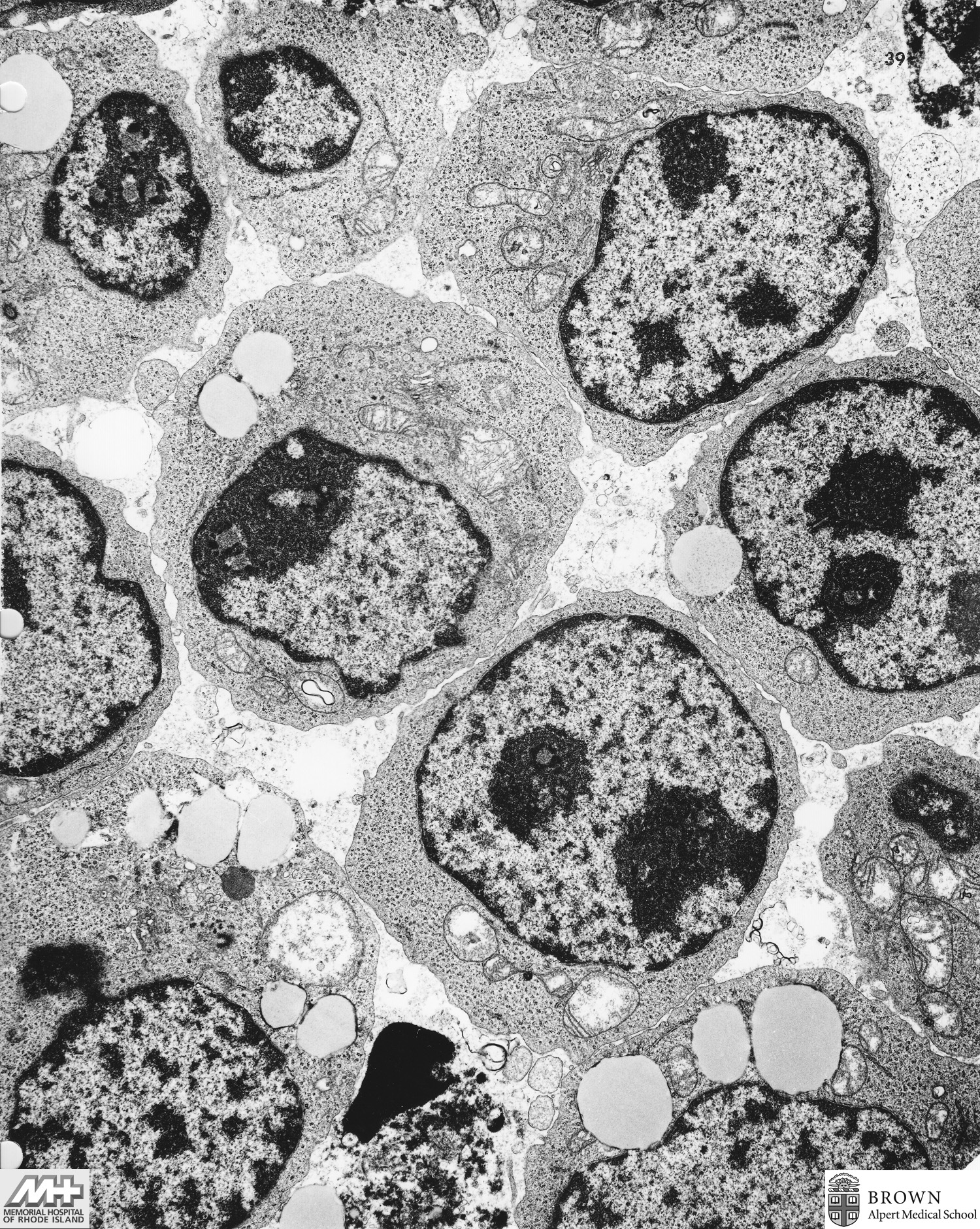
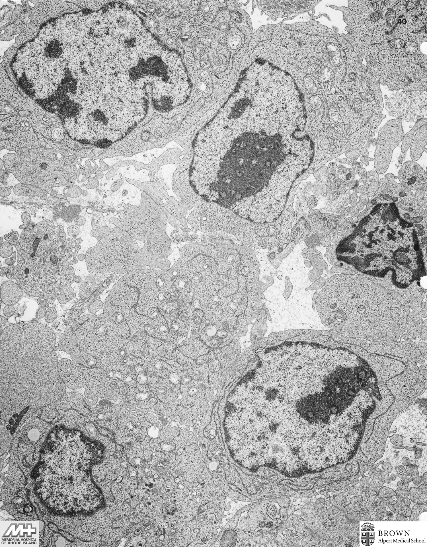
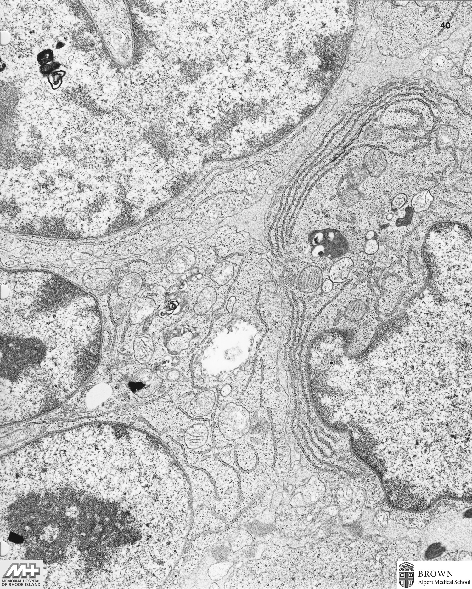
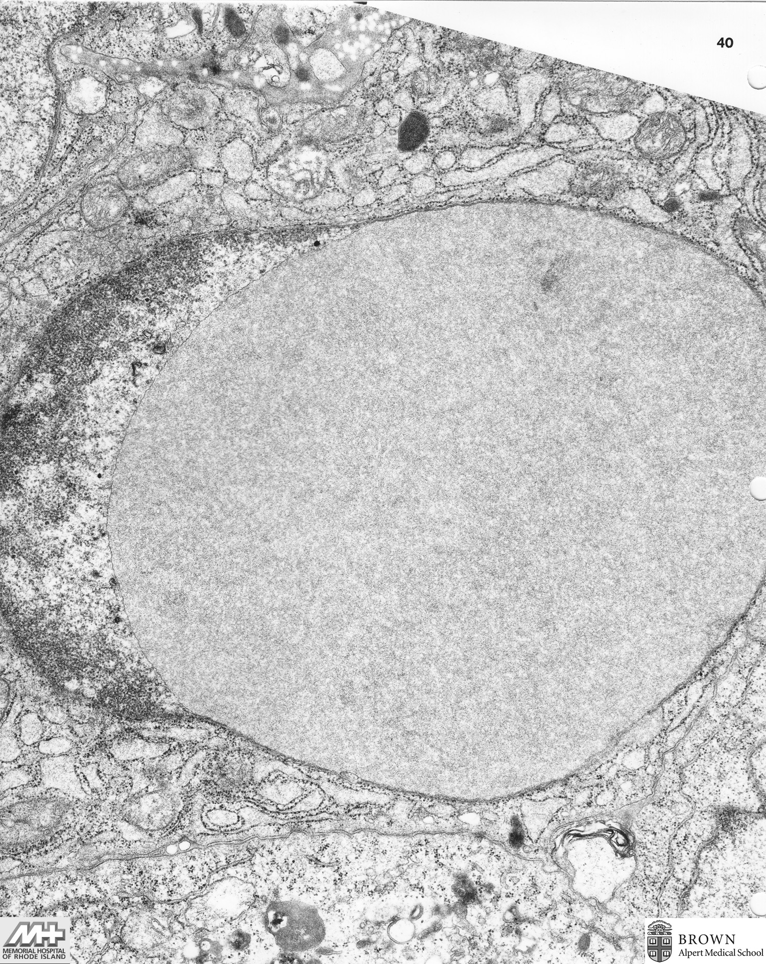
Case 37: Small non-cleaved lymphoma (Mediastinal lymph node)
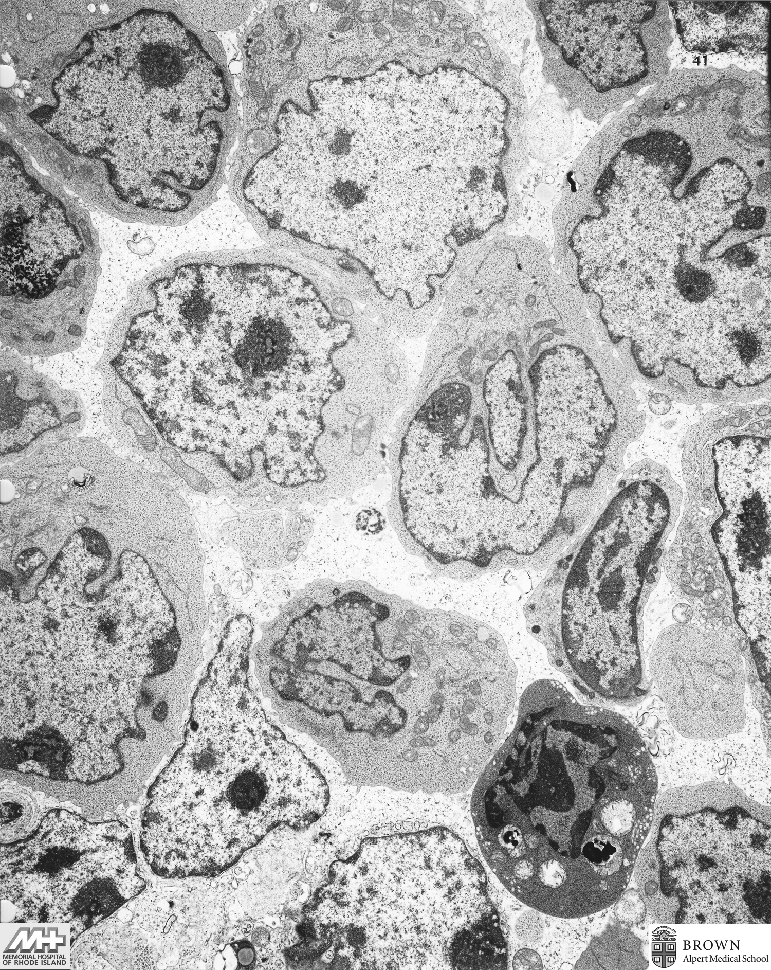
Case 38: Immunoblastic lymphoma (Cervical lymph node)
Case 39: Diffuse large cell lymphoma (Lymph node)
Case 40: Metastatic breast adenocarcinoma (Lung)
Case 41: Endothelial cell factor VIII, gold stain (Blood vessel)
Case 42: Anemone cell tumor, Transtional cell carcinoma vs. Mesothelioma (Peritoneum)
Case 43: Yolk sac tumor (Mediastinal mass)
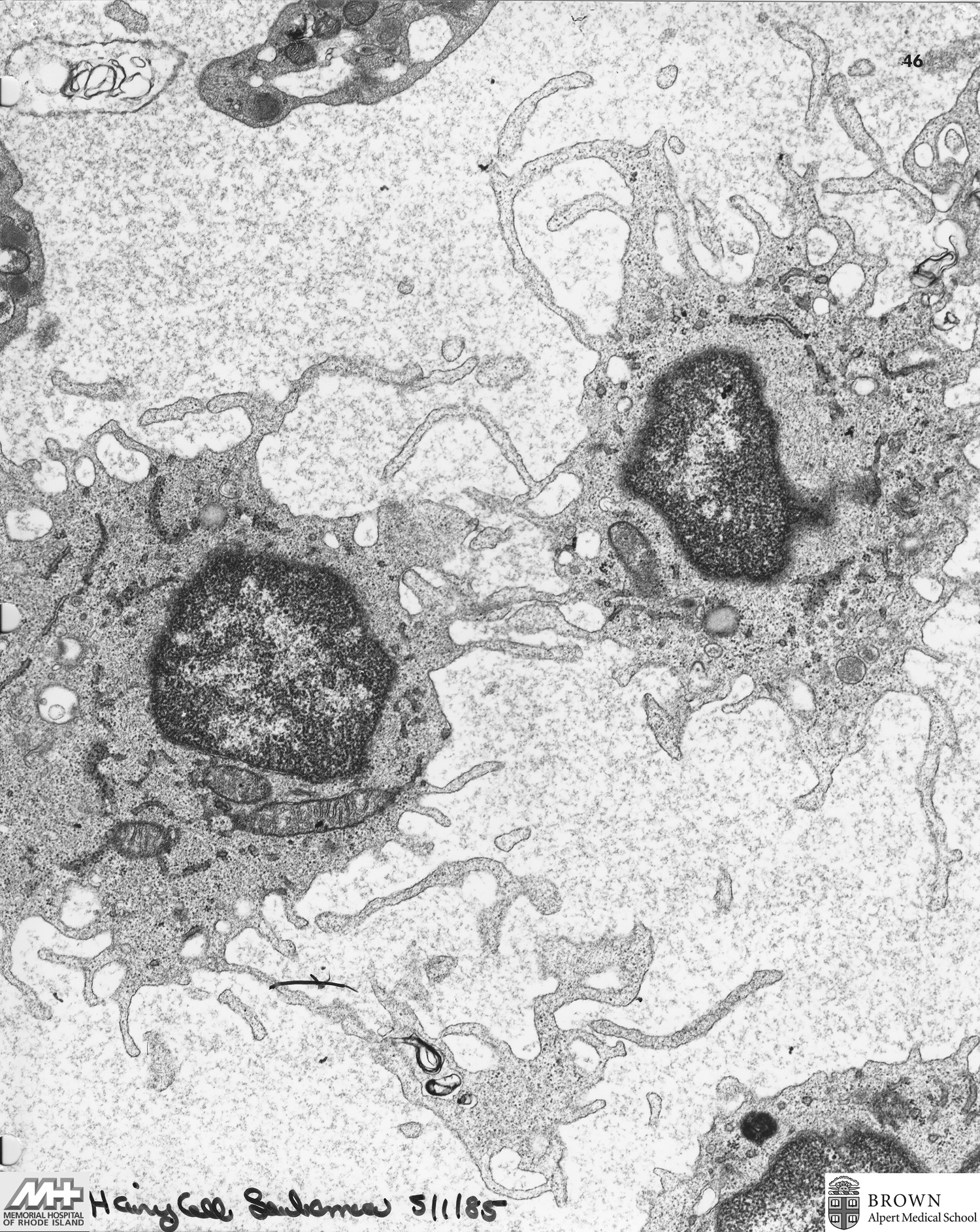
Case 44: Hairy cell leukemia (Blood)
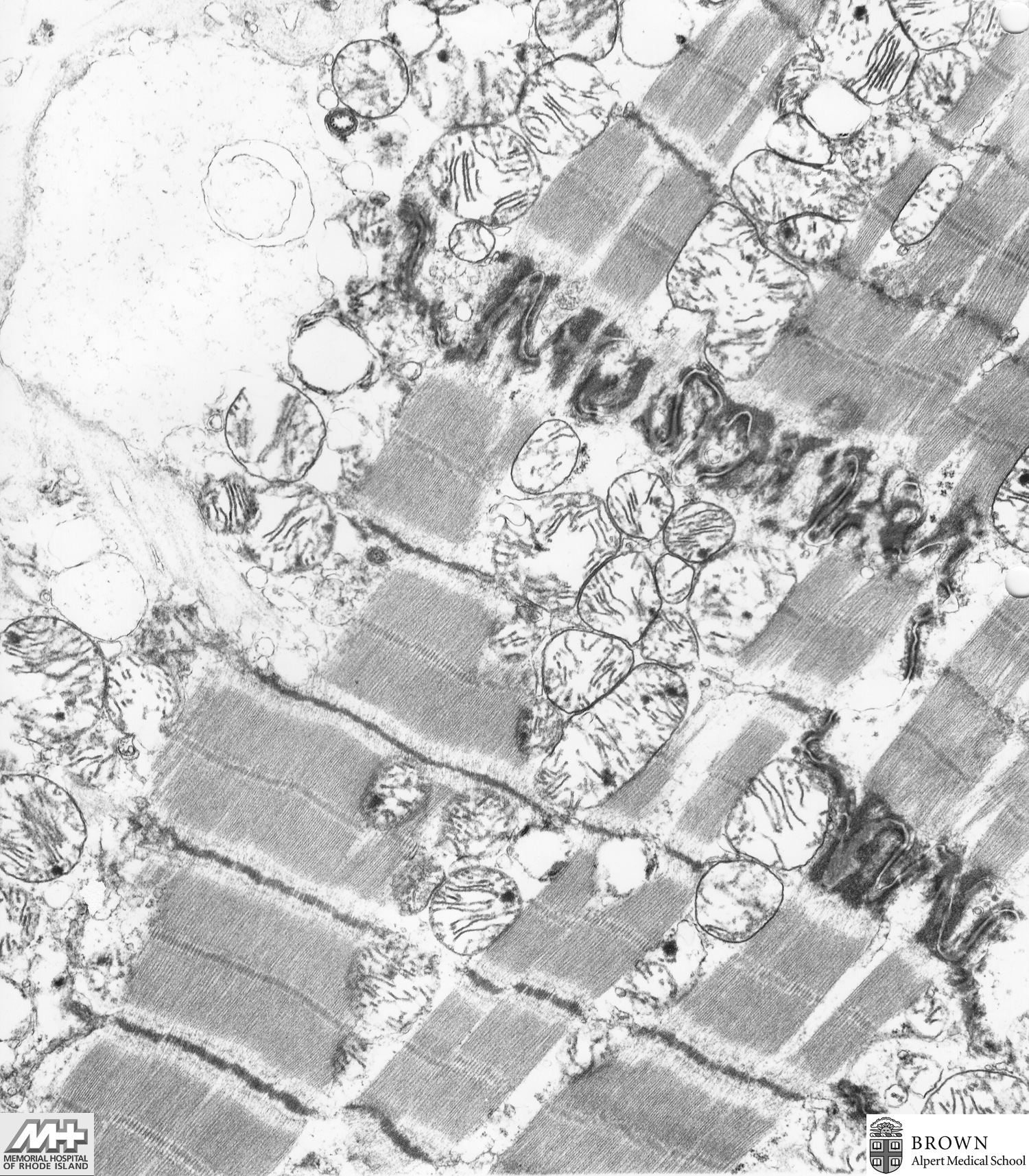
Case 45: Heart (Normal)
Case 46: Clear cell carcinoma (Ovary)
Case 47: Infiltrative ductal carcinoma (Breast)
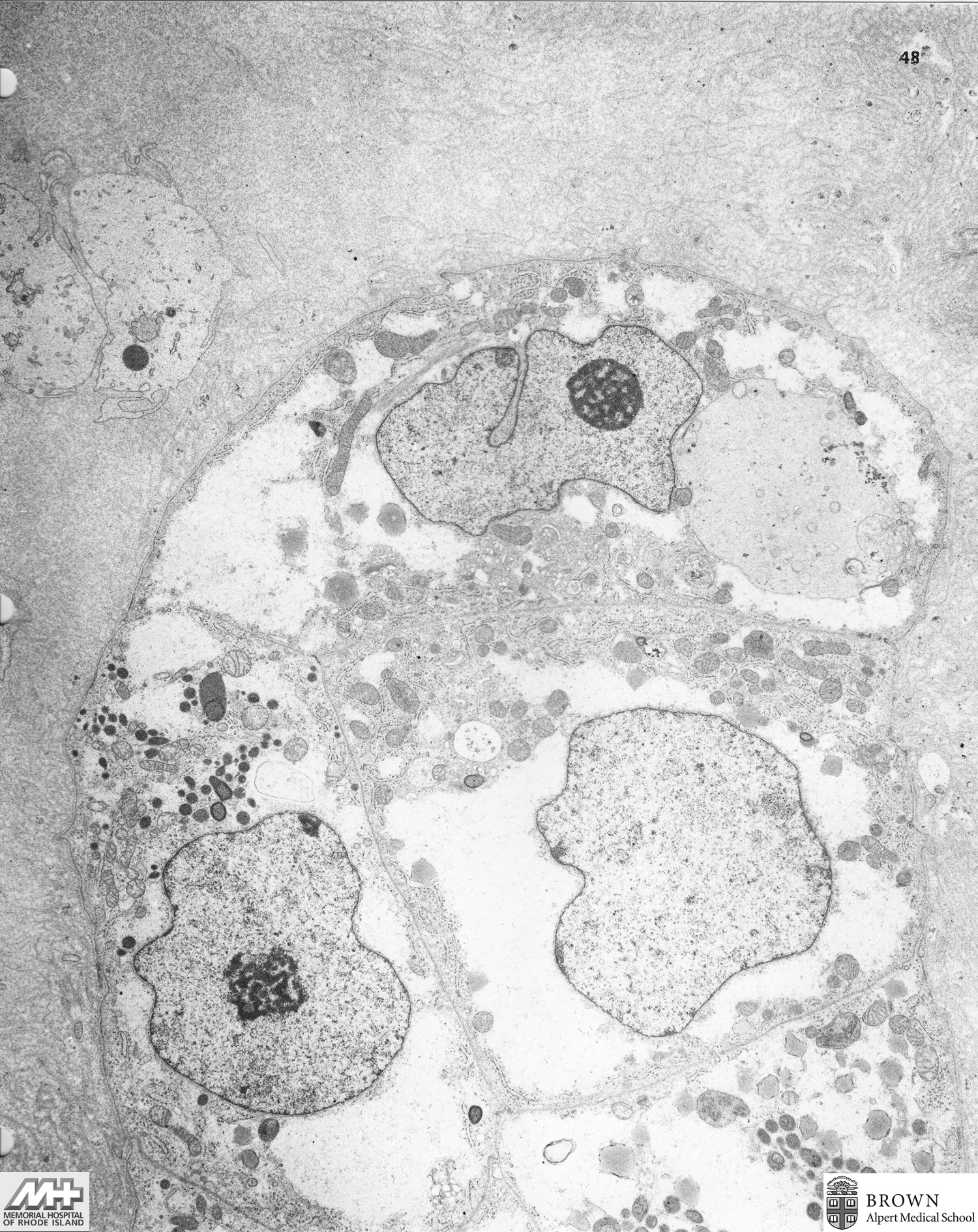
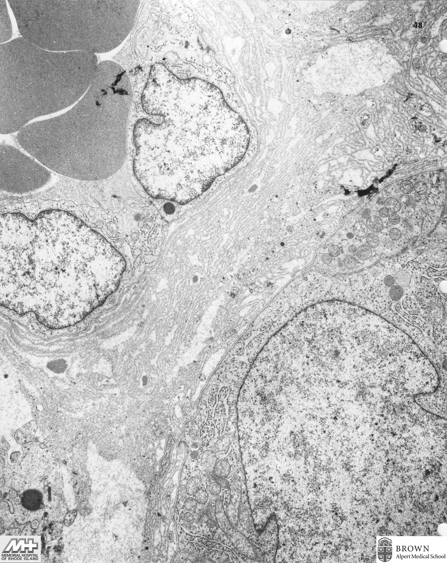
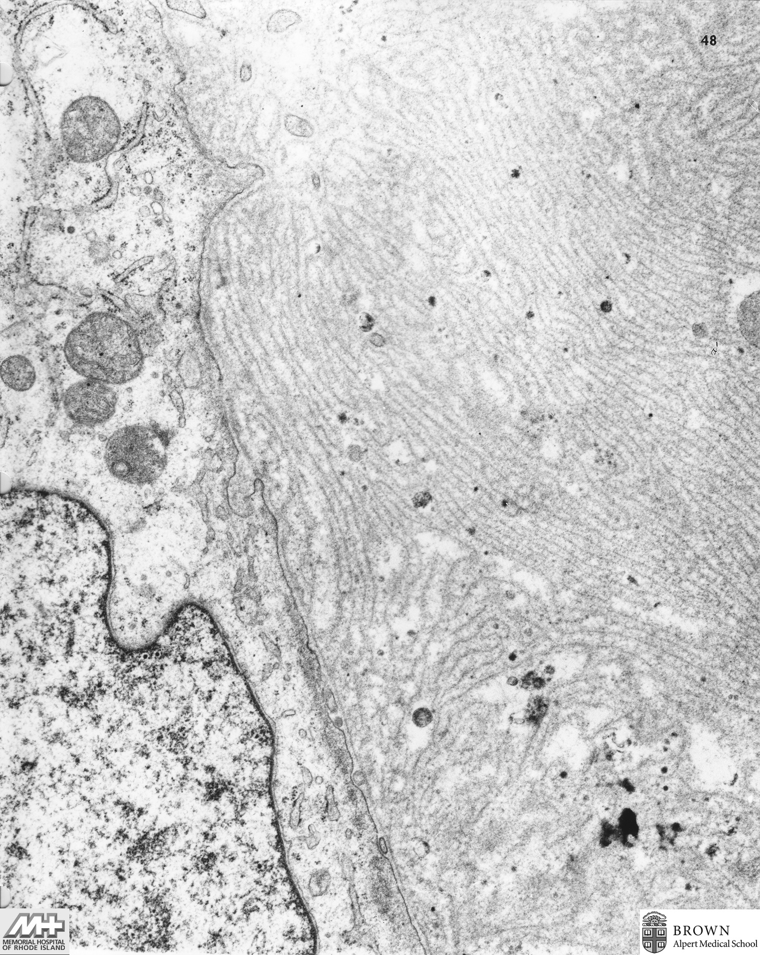
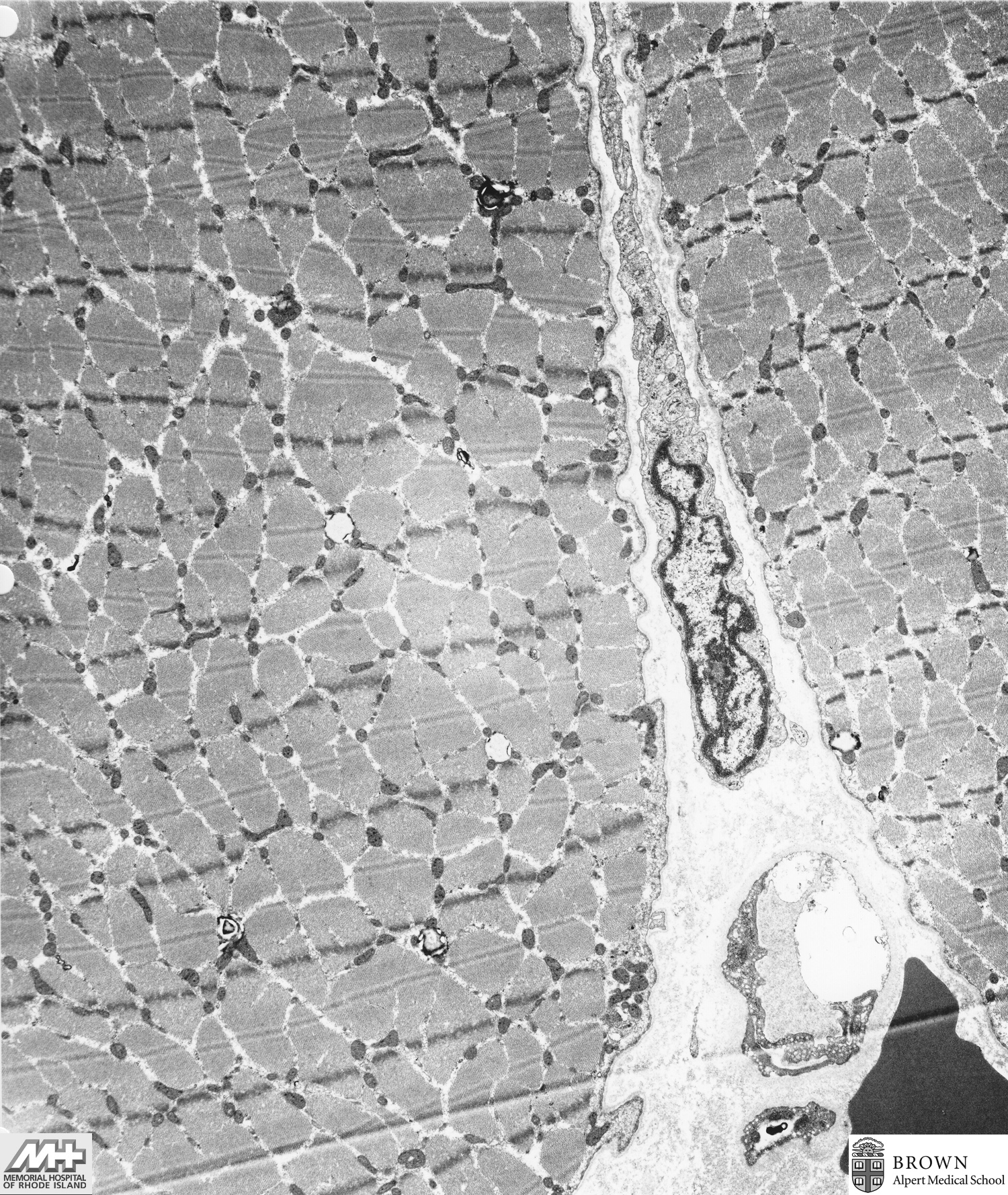
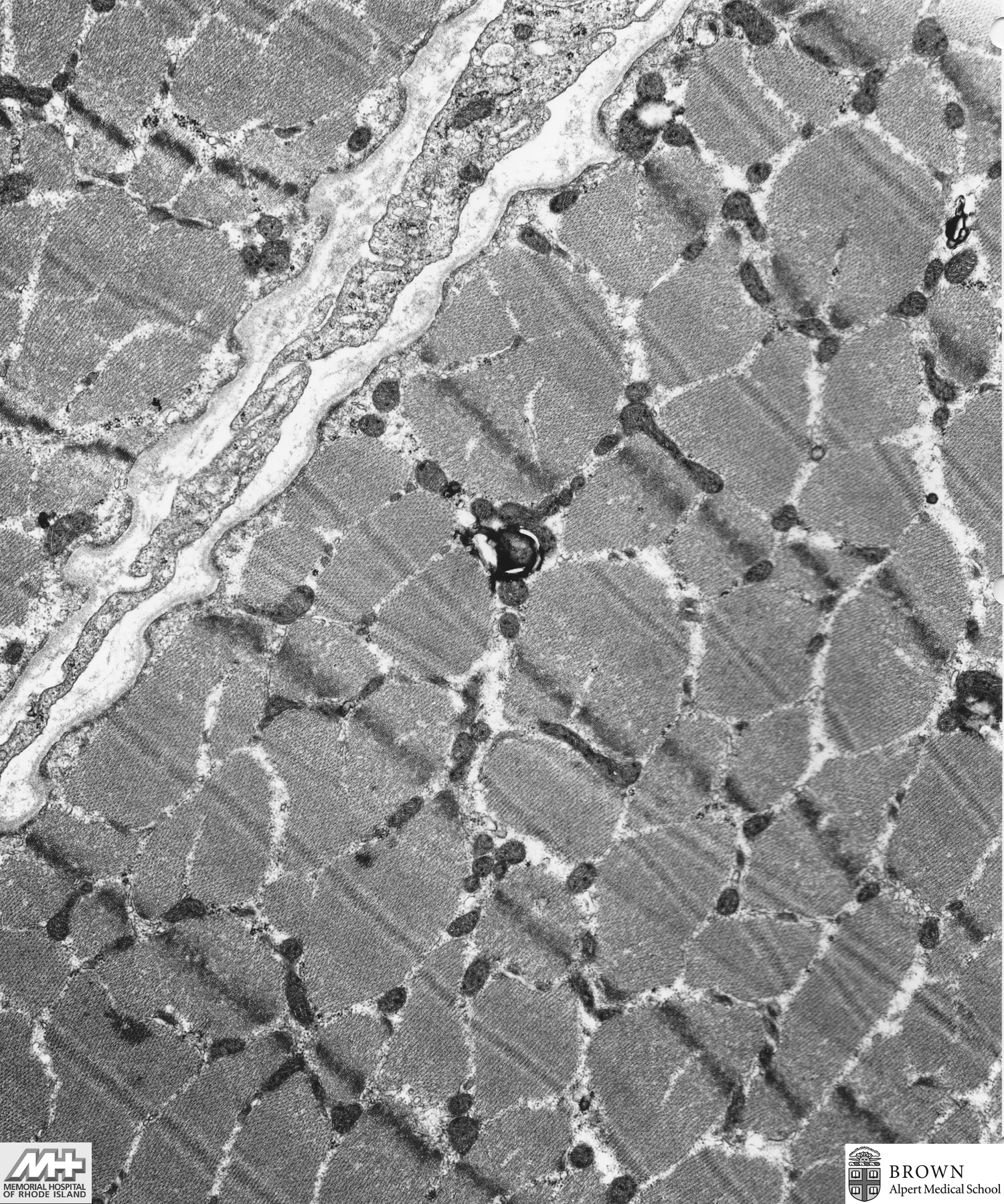
Case 48: Type II and minimal Type I fiber atrophy (Quadriceps biopsy)
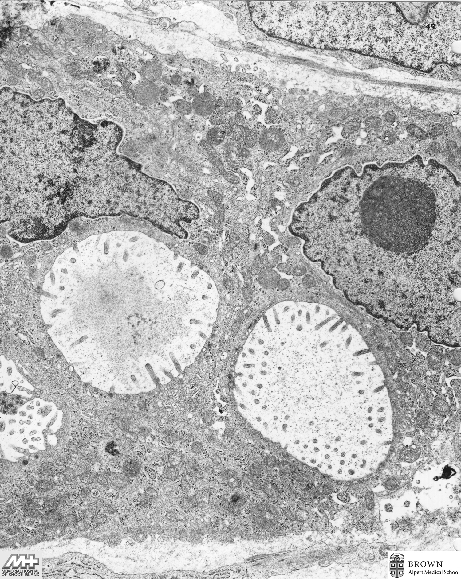
Case 49: Metastatic amelanotic melanoma (Groin lymph node)
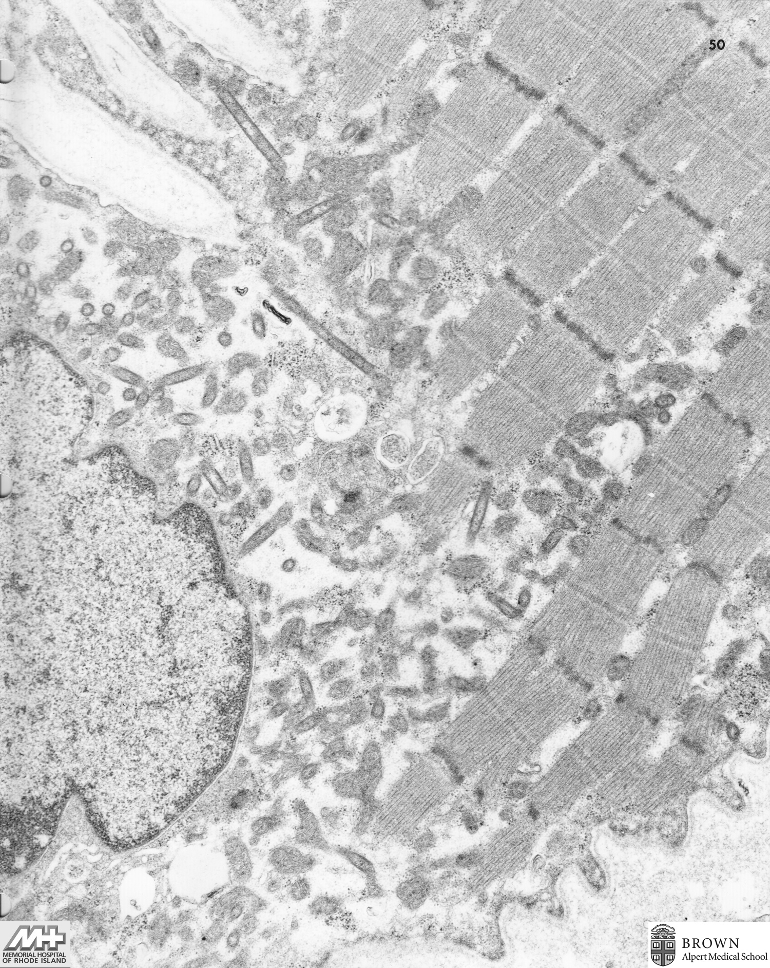
Case 50: Carcinoid (Small bowel)
Case 51: Leiomyosarcoma (Retroperitoneum)
Case 52: Muscular dystrophy vs chronic myositis (Quadriceps)
(Scanned and organized by Shaolei Lu, MD)