Aspergilloma
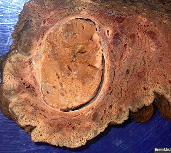
This is a specimen from a right upper lobectomy. The patient was a 75 year old man with a chronic cough.
Routine chest x-ray showed a 3 cm right upper lobe mass. Bronchoscopy x 2 and CT directed fine needle
aspiration were negative for tumor. The mass with approximately 2 cm greatest diameter represents a fungus
ball due to the growth of aspergillus colonies. The cavity may be lined by epithelium which often shows
squamous metaplasia. The contents of the aspergilloma include colonies, blood clot, and cellular debris.
Contributed by Dr. Michael Klein
1 minute clinical correlation