Measuring eye movements
Reading: RHSC2 Appendix 1
By "eye movement" we mean rotation of the eye with
respect to the skull, whether the skull is moving or not. Just rotating the
neck to "move" the eyes does not constitute an eye movement, but does
change gaze.
Eye movements are the bioengineer's research arena par
excellance
David Robinson of JHU, Ed (left-handed) Keller, UC Berkeley;
the instrumentation, particularly for monitoring eye movements, favors engineer
researchers
Specifications for an ideal instrument
Goals: accuracy, sensitivity, bandwidth = speed,
range in degrees. doesn't interfere with vision or head movement, is not visually
distracting. has electronic readout. can be done in the dark. Must the head
be clamped, or does the ideal system ride along with the skull?
For RHSC, the important criteria is noise as a function of
frequency. Low frequency noise disturbs position, but not velocity, measurements.
Internal
monitoring of eye position
The brain knows where the eye is looking, even in the dark, by monitoring
the output of the motoneurons controlling the eye muscles. Since the muscles
can't fatigue and never carry loads, the motoneuron signal OUTPUTS are always
faithful versions of eye position. Yes, some visual calibration must have gone
on in the past, but presuming that is all okay, no proprioception is needed.
Direct Viewing
Clinical. Can you resolve 1 degree saccades by direct viewing? yes. Observe
smooth following. Observe vergence.
TV
Making videotape of eye movements is the next step: what are the limitations?
no electronic output, 30 Hz update frequency from TV. The camera may occlude
parts of the visual field. Scan paths for watching people look at advertisements.
the limitations of TV sampling rate for monitoring saccades
Photoelectric & infrared
Measure reflectance of IR from sclera vs iris. The
limbus is the region of transistion from the cornea to the sclera (Oyster, p.
78) The method looks at the sclera/iris boundary and will definitely
provide an electronic output of horizontal eye movement.
DEMO: See
the ASL 210 using infrared transmitters and detectors:
an arrangement of differential recording. The limbus is sclera-iris boundary.
Demo: ASL210 eye movement "scanner"
Pattern recognition of the boundary-the demo
detect the Corneal/scleral boundary
limbus tracker: ASL 210
Need banana to BNC adaptor: connect to digital storage scope.
Study Horizontal eye movements only to begin with.
Suggested settings: Use left channel only.
time base of scope: 200 msec / div
gain on ASL 210: about 1.0, on the scope, 200 mv / div
turn Crosstalk all the way CCW.
Set Linearity at midpoint.
If pure vertical up-down movements are made in the horizontal mode, the extent
of signal change is called "crosstalk" and adjustment of crosstalk
and linearity knobs may be in order.
Observe saccade and smooth
pursuit eye movements on the scope.
Attempt calibration.
Show usefulness for juggling
measurements, and see if vestibular nystagmus can be demonstrated.
Demonstate that the ASL 210
works fine with the lights. off.
Attach the digital scope
and watch the trace with eye only and head only movements. Try to make smooth
pursuit on a stationary background. Show how slow vergence is.
Reflection
Reflection off the cornea, the various Purkinje images from the cornea and
the lens:
Use the a low-energy laser beam to reflect off the cornea...
 ?
?
is the equation for deviation, where alpha is the position on the screen and
theta is the eye movement. d is the diameter of the cornea, or the small spherical
target on the surface of the eye.
The
RIT corneal eye tracker
topic: Feedback for stabilized images: See previous lecture, on opening the
loop for optokinesis.
Barlow: A small diameter reflecting sphere on the eye...a drop of mercury!
Electrooculogram EOG
EN123 Lab
"A" : circuit with UGVF and long time constant electrode interfaces
(HP filter) then an instrumentation amplifier; then a LP filter at an op amp.
Source of potential: the continuously flowing current from the outer segments
to the cell body of the rods and cones of the retina. More current in the dark.
Does the EOG change when the eyes are closed?
various artifacts from muscle...
see Mcgill
website about EOG and caloric testing experiment.
Go to the Biological Signals "book" icon and look at the EOG
pages!
Search coil
Mutual Induction and eye movement measuring:
need to have a sinusoidal time-varying B-field so that steady changes in gaze
result in steady RMS changes in EMF.
A Helmholtz coil provides a relatively uniform (spatial) magnetic field through
the eye.
Imagine a coil of wire wrapped around the eye, tucked under the eye muscles.
What is the voltage of the eye coil leads?

E is the emf, or electromotive force, or voltage. what is F? Flux.

where a is the surface normal vector of the coil area. For the units
of volts, B should be in Tesla (one tesla = 10^4 gauss) and area should be in
meter^2. The units of flux are webers, in that regard. Tesla-meters squared.
So if we make B change with time then even when the head isn't rotating
a nonzero emf will appear. Use phase-locked loop to monitor the eye movements
as a function of change of angle of a with respect to B. Need to take
account of the number of turns of wire under the eye muscles.
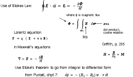
In "electromagnetic recording" Helmholtz coils will be around the
head, the other coils around the eye: fine wire placed at the equator, under
the eye muscles, with the ends of the wire led out to a connector. Griffiths
discusses mutual inductance in chapter 7. Mutual inductance is the term RHSC2
uses in describing "electromagnetic recording."
How can the search coil give a sustained reading to a new fixation? Have the
B field be time varying: sinusoidal. In fact two different B fields of two different
frequencies can influence the coil around the eye, and both left-right and up-down
movements can be monitored "simultaneously."
Example: Assume you have a high power stereo amplifier (PA system?)
with a "cosine" wave generator set to 1000Hz driving an 8 ohm pair
of coils (Helmholtz coils) to produce a 100 gauss p-p B feld through the eye.
(What is the relationship between current and B field? B = m0
NI for a solenoid) If the diameter of the eye is 2 cm and it's wrapped
10 times with 30 gauge wire, what is the p-p voltage output of the coil when
the plane of the coil is perpendicular to the B field?
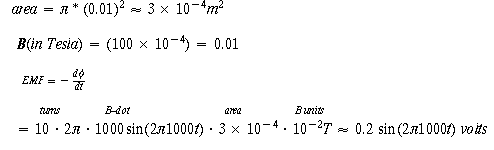
where Hz are converted to radians and gauss to telsa and cm to meters. output
is in volts.
What would be the voltage with the eye turned 45 degrees?
Use of Helmholtz coils to produce spatially uniform B field. ETS-Lindgren
Corp. & a link
from Univ of New Mexico
Ultrasound images of the eye: for seeing changes in lens shape.
Summary
* specs: speed, accuracy, lack of interference
with vision, measure in the dark.
* Direct viewing, TV, self-report of fixation
* Photoelectric--infrared
* Reflection, off the cornea
Stabilized vision
* EOG, with electrodes
* Search Coil
~ methods for measuring pupil diameter and lens shape