Vestibular Influence on Eye Movements
Reading
RHSC2, Chapters 2, 9.2
Victor J. Wilson & Geoffrey Melvill-Jones, Mammalian Vestibular
Physiology, Plenum Press (1979) try chapter 3. [QP471/W54], on reserve
The Neurology of Eye Movements 2nd Ed., by John Leigh
& David Zee, F.A. Davis Co., Philadelphia (1993). RC 321.C66
Lisberger & Sejnowski, Nature 360: 104, 159;
"Motor learning in a recurrent network model based on the vestibuloocular
reflex," (Nov 12, 1992)
Vestibuloocular reflex
The most important influence of the vestibular apparatus on eye movements
is the vestibuloocular reflex (VOR). The VOR is a compensatory
eye movement to cancel head rotation and maintain fixation. As you learned in
the Canal Mechanics lecture, the dynamics of the SCC are such that the vestibular
apparatus is fundamentally an angular accelerometer that acts as a velocity
transducer in the physiological range of sinusoidal rotations. Thus the VOR
is not a sustained reflex; after many seconds of steady rotation the stimulus
for eye movement will die down. Furthermore, during continuous rotation in one
direction the limit of compensatory eye movement will be reached after 50 degrees
or so; after that a vestibular nystagmus (VN) will commence;
VN has a fast and slow phase; the quick phase will reset the eye to begin another
slow phase of true VOR. The VOR can be measured in the dark, and in fact should
be; in the light another factor, optokinesis, (and its attendant
nystagmus) competes with VOR for control of eye movements.
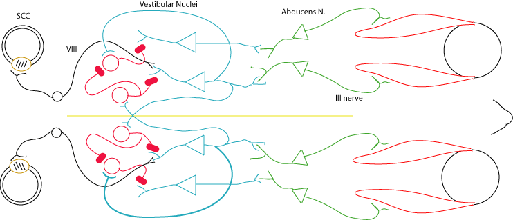
Vestibular nucleus
The star of this lecture is the vestibular nucleus: its afferents, its efferents,
and its role in control of eye movements. In fact it's nuclei plural: there are
several (4, + x,y,z nuclei...) on each side of the brain. We'll mainly concern ourselves
with the parts of the vestibular nuclei which receive afferents from the horizontal
canals, and how the signals from the horizontal canals carry out the vestibuloocular
reflex, in the horizontal plane.
"The vestibular nuclei provide the largest single source
of fibers to the oculomotor nuclei..." RHSC2 p. 218, chpt 8.
The Vestib Nuc is also a major source of input to cerebellum.
Vestib Nuc affects various spinal reflexes associated with balance.
Inputs to Vestibular Nucleus
Other inputs to Vestib Nuc: from cerebellum (flocculonodular node) and the
deep cerebellar fastigal nucleus. AND from contralateral Vestib Nuc! See RHSC2
chpt 9.2, p. 216:
Vestib Nuc also responds to vision: visual slip, so it must receive
a visual input.
The Vestib Nuc also needs eye movement information, but not from
proprioception: RSHC2 p 217: "Since it also receives connections from both
of the main accessory oculomotor nuclei--the interstitial nucleus of Cajal and the
interstitial nucleus of the medial longitudinal fasciculus (MLF)--from the preoculomotor
reticular areas, and from the oculomotor nuclei themselves, it may well be the point
at which the eye movement information that is present in vestibular nuclei
activity is added to signals carrying visual and vestibular estimates of head velocity..."
superior vestibular nucleus = SVN; medial vestibular nucleus =
MVN;
�9.2 of RHSC2. "...MVN neurons respond mostly to stimulation of the
horizontal canal."
Cell types in the Vestib Nuc
The responses of neurons in Vestib Nuc, p. 222
type I: ipsi canal drives it, 2/3 are type I
turning to the ipsi side is excitatory...
type II receives input only from type I on the other side of the brain, and
are inhibitory to type I on the same side. It seems that type II's also project
to abducens nucleus...
Leigh & Zee p. 22: "...ipsilateral type I vestibular neurons drive
contralateral type II neurons" See Fig.'s 9.10 & 9.11 in RHSC2. Type
II neurons can inhibit other Vestib Nuc cells!
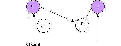
A push-pull system, to convert the unidirectional into bidirectional response.
Shown below is half of a push-pull arrangement; the right side type I must
have a symmetric connection to the left...
p. 220 "The major descending outflow from the vestibular nuclei is to the spinal cord,
in the vestibulospinal tract (originating in the LVN, which is somatopically arranged),
and in the MLF (from the MVN). The LVN projects to all spinal levels and probably
influences muscle tone and posture, whereas the MLF projects only to the cervical
area (very likely serving the vestibulocollic reflexes)..."
RHSC2, �9.2.4 starting on the bottom of page 221. 66% of Vestib
Nuc cells are type 1, which respond to ipsi canal stimulation, 33% respond to
contra.
Static tilt
Involvement of utricle and saccule. More pronounced in rabbits. See Figure in
RHSC.
Blow up a balloon and draw eyes on the side. Draw vertically slit pupils, for orientation.
Put on bunny ears. Rabbit makes "pitch" rotation: one eye looks up, the
other looks down.
Make rabbit look down. Show how torsion movements would maintain the gaze of the
rabbit. Does looking up and down evoke static tilt in humans?
Dynamics
Gain of VOR. Ideally, the gain of the VOR should be precisely
-1.00, to compensate exactly for head rotation. What is the gain of the VOR in
the dark? It seems to be a little less than 1.0. And we know from previous
work on the canal, that the magnitude of the vestibular response to a ramp will
decline as the head rotation continues. Why not measure VOR gain in the light? If
you do, results will be mixed with optokinesis.
Use of guinea pig for consistent VOR.
Modelling the VOR: See diagram below:
Startling observation for model: The time constant of decline
for the VOR is greater than the time constant of decline for the vestibular system
recorded in VIII neurons! "Velocity storage" needed, at the level of the
Vestib Nuc, in association with the cerebellum.
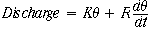
Latency of VOR: short! about 15 msec! Why? partly
because of the speed of the hair cell response.
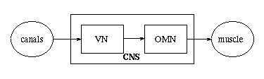
What does a motoneuron in the Oculomotor nucleus (OMN) do?
this "force" overcomes the "mechanics of the plant"
Frequency response & Demos
Why can you read better when your head is rotating, vs
the page rotating?
How to measure VOR-gain, phase
From Leigh & Zee, page 21: "For each of the vertical canal-extraocular muscle pairs, one
muscle receives a crossed and the other an uncrossed innervation."
What to say about frequency response? Fig 2.21 of RHSC2 shows a "flat curve" out to 1 Hz.
Ellen Barton's thesis says it's flat out to 4 Hz. This is the "physiological range" for head
rotation. What do you get with the Bode plot sketch of the vestibular transfer function, as
you learned in Circuits class? Don't forget the s2 term.
What about phase change? See RHSC2 chpt 2 figure.
Caloric testing of the canals
Lee & Zeigh, pages 28-29: while supine and head tilted 30�
up, infuse 45�C = 113�F hot water into external auditory meatus for 40 sec, and
then after a recovery period, at 30�C for 40 sec. thermal gradients principally
stimulate the lateral semicircular canals... Maximum slow-phase velocity is generally
considered the most reliable index of peripheral vestibular function. Observe vestibular
nystagmus with no head rotation!
2006: Misha says use cold water... will
cause nausea as well as nystagmus!
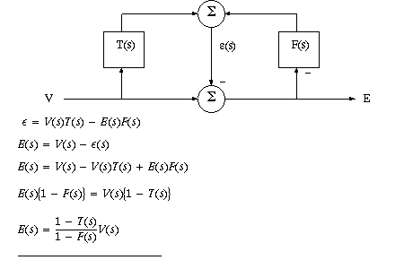
Lisberger & Sejnowski model
and see Sejnowski + Churchland book...
The diagram for the Lisberger & Sejnowski system is shown below. It's a positive feedback
system that remembers time constant mismatch. If T(s) = F(s) then E=V
Cooperation with optokinetic nystagmus in the light
Gain of OK reflex should be +1.00
OK reflex comes on slower than VOR, but doesn't taper off. The two effects complement
each other. What transfer function models the slower OK? First order LP...
When both OK and Vestibular input are active, why don't we get a gain greater than 1?
Or a gain almost zero?
Review of Wilson & Melvill Jones
Chapter 5, "Labyrinthine input to the vestibular nuclei and reticular formation"
good figures 5.7, 5.8 for contralateral stimulation...
p.147 Vestibular neurons are typically spontaneously active
p. 150 cell types, see above...
158-160 visually generated signals, 1975 ref, UCB work.
fig. 5.12, eye-movement related units (not responsive to head only movement)
Fig. 5.8. Commisural pathways for inhibition: type I drives type II across the commisure,
then type II inhibits local type I.
Chapter 7, "The vestibulospinal system"
neck reflexes
Chapter 8, "The vestibuloocular system"
pages 273-80, nystagmus
Is it an oscillator?
Is a threshold involved? look at tracings...doesn't seem to be
role of reticular formation
Minimal, but realistic, neural model "wiring diagram"
for VOR
(2006) Shown below is a minimal model for horizontal VOR,
from the SCC's on the left to the eye muscles on the right. The model is a top-down
view. In green are the OMN, with the Abducens nuclei responsible for controlling
the lateral recti. In red are inhibitory neurons in the Vestib Nuc, as well
as "projection cells" in blue from the Vestib Nuc to the OMN. One
"design spec" met by the model: that either SCC by itself can influence
all 4 eye muscles. We know from monkey studies, where one VIII is inactivated,
that after less than a month the remaining SCC can "reprogram" its
synaptic weights (gains) and restore reasonable VOR.
Notice that the medial pathways in between the Vestib Nuclei
on both sides form a push-pull system: a positive feedback loop with two inhibitory
neurons.
The lateral pathway blue projection neurons of the Vestib
Nuc must be spontaneously active because in the minimal model they have only
inhibitory input. If the contra canal hair cells depolarize and excite the corresponding
VIII nerve, the contra projection neuron drives an inhibitory cell that inhibits
another I cells that normally suppresses the ipsi projection; the result: the
ipsi lateral rectus contracts.
How does the ipsi SCC control the ipsi lateral rectus?
By the local feedback pathway from the medial projection neuron in the ipsi
Vestib Nuc to the lateral red inhibitory neuron, shown in thick line, above.
Where are the inhibitory neurons seen by anatomists? In the
Vestib nuc., and possible type II cells in abducens nucleus.
For instance, how is the ipsi lateral rectus inhibited during
VOR? See RHSC2: page 218. Connection of Vest. Nuc. to abducens: "In the
case of the abducens nucleus, single shocks of the ipsilateral vestibular nerve
(VIII nerve) produce inhibitory postsynaptic potentials, whereas during repetitive
stimulation the abducens neurons show periodic electrical phenomenon in step
with the resultant horizontal nystagmus."
In the VOR, a head rotation to the left causes the left MR to contract. The projection to the
MR is "ipsi," from OMN, cranial nerve III. Therefore the left horizontal canal, and left VIII
nerve, must be excited by head rotation to the left. [Looking down from the top, CCW
rotation must depolarize the hair cells of the left horizontal canal.]
The abducens, driving the right LatRec, is uncrossed and on the other side. To
contract the LR on the other side the VN sends a projection across to the other
Vestib Nuc (type II units) which then drives the abducens.
The left Vestib Nuc must excite the left OMN which contracts
the left MR. The right MR is relaxed by the reduced activity of the right canal
hair cells.
What about the other eye? the right LR must contract.
But the right VIII nerve has a complementary response to the left VIII nerve...its
activity decreases. [Decreased activity in the right Vestib Nuc causes "relaxation"
of the LR antagonist, MR, on the right...] However, an excitatory signal
from the left side, it turns out, crosses over to stimulate the right abducens
nucleus. See RHSC2, Fig. 9.7. The Horizontal canal zone of Vestib Nuc inhibits
ipsi abducens units, excites contra abducens. Is there a crossover directly
from one Vestib Nuc to the other? Yes. Type I neurons in one Vestib Nuc cross
over to stimulate type II neurons in the opposite Vestib Nuc. See figures on
RHSC2 p. 223. [ Not to be confused with the type I and II hair cells that RHSC
describes in chapter 2; (misprint for their label in figure 2.5.)] In engineering
terms, this is a push - pull system.
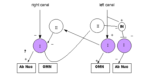
How does the lateral rectus on the left become inhibited? There may be
an inhibitory interneuron from VIII to the type 1 cell projecting to the abducens
nucleus, AND there may be a projection across the commisure from the right Vestib
Nuc, to a type 2 cell that goes to a IN neuron that projects to the type 1 going
to abducens. Both paths would give another push pull effect on this other pathway.
See 3 negatives in the path across the commisure. Path across the commisure
is disrupted if one VIII is injured.
Yes, there must be a second inhibitory neuron on the excitatory
neuron going to the abducens nucleus. And the reduced type I activity on the
contra side, plus the increased inhibitory activity from the ipsi side must
reduce the activity of the inhibitory neuron riding on the type I projecting
to the abducens nucleus. There's gotta be some way to reverse the sign . There
are other possibilities, in principle, but the 2 inhibitory neurons is a valid
model to account for the whole VOR, from the horizontal canals.
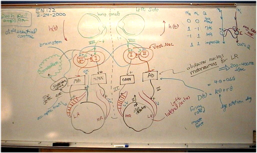
See whiteboard image for a year 2000 "EN122 historical" diagram
of VOR control, with an extra excitatory input in the pathway to the abducens
nucleus.
Quick phase of Vestibular Nystagmus
What about the quick phase of vestibular nystagmus?
Researchers can't say for sure what causes quick phase to be triggered.
Quick Phase VOR does "suppress" the normal VOR response.
RHSC2 Chapter 2, "...even complete removal of the eye and muscles from the head does
not abolish the rhythmic (nystagmus) activity of vestibular nystagmus...in motoneurons"
What does this imply about mechanism of nystagmus?
RHSC2 p.228: something must "build up" (or a threshold must be reached) to generate a
quick phase saccade...
during quick phase the opposite set of muscles must come into action...the
normal VOR circuit above must be overridden by a saccade control .
"About half the units in the vestibular nuclei of the alert monkey respond to spontaneous
saccades or nystagmus quick phases by bursts or pauses around 15 msec before the
movement itself."
The MLF: quote of p. 229. Does its abolition remove the VOR? no, only the fast
components.
Head and eye movement & VOR, fig 2.22. Slow phase is VOR.
"override" of VOR...try it with a mirror angled
in front of one eye
SUMMARY
* VOR is fast open loop response for stabilizing vision
vestibular vs optokinetic nystagmus.
* Compare VOR response to VIII nerve response: longer time constant
* Anatomy of VOR, excitation & inhibition to MR & LR on both sides
(Push pull system)
* Physiology of VN: type I (ipsi) and type II (contra-driven)
* too much rotation causes nystagmus...then nothing
* OK response "compensates" for long-time rotation
* neural integrators & freq. response
-----------------------------------------------------------------------
EXTRA MISCELLANEOUS NOTES:
what about OKAN and V after nystagmus?
JNP 49: 134 (1983) Paige.
Describe physiology of Vestib Nuc neurons?
SUPPRESSION OF VOR
by quick phase of nystagmus
by visual fixation during head turning
Q: neck proprioception & PREDICTION,
FROM SELF-ROTATION... keep head still, move body...not much reflex... static tilt
from otolith organ example with balloon-rabbit, showing that superior recti (oblique?)
must be active... Q: Continuous rotation produces nystagmus part VOR, part override
of quick phase what happens if rotation goes on for, say, 30 sec? " "
" stops? MEASURE IN THE DARK, AND IN THE LIGHT...OKN the other kind of nystagmus...starts
up slowly, but continues indefinitely. canals provide velocity signal, OMN needs
position output (or do we?) Q: implies what? "neural" integrator Frequency
response, rolls off at 2-6 Hz stimulate with back-and-forth rotations...has acceleration
components how to reconcile with vestibular frequency formula s*s/(s+a)(s+b) The
muscle plant has an "integrator" built in probably different from velocity-to-position
integrator... Anatomy of inhibition and excitation HC=horiz canal ipsi rot + type
I, - type II (type II is inhibitory...) type I drives ipsi MR excitatory projection
of type I to contra type II How does contra LR get excited? ipsi Vestib Nuc excites
contra abducens... COMBINED HEAD AND EYE MOVEMENTS what about fast phase of V-nystagmus?
build-up of inhibition... other overrides, for smooth pursuit KEC: VOR capable of
stabilizing gaze over a remarkable range of frequencies (0.05-14 Hz) and velocities
(up to 350�/sec) of head rotation. chapter 2: We've consider some of this material
in a previous lecture on the vestibular apparatus; we worked up to understanding
the frequency response (dynamics) of the system. There is a basic 3-neuron open-loop
reflex arc, with a delay of 15 msec (vs 150 msec for smooth pursuit) Above 2 Hz
the VOR gain drops below 1
NYSTAGMUS-a beating back and forth in response
to unidirectional rotation-vestibular, slow and quick phases. vs optokinetic nystagmus.
QUESTION: how long can vestibular nystagmus
continue? 30 sec? see homework problem...