Notes on cerebellar circuitry
----------------
Reading:
John Nolte: The Human Brain: An Introduction to Its Functional Anatomy, 5th
Edition, Mosby (2002) chapter on the cerebellum.
JF Stein, Functions of the Brain, Oxford Univ Press (1985)
W. Nauta and M. Feirtag, Fundamental Neuroanatomy W. H. Freeman &
Co. (1986)
Maseo Ito: The Cerebellum and Neural Control, Raven Press (1984) QP379
.I86
unorganized
archive en122 notes on cerebellum 1988-98
Websites: links valid 3.2005.
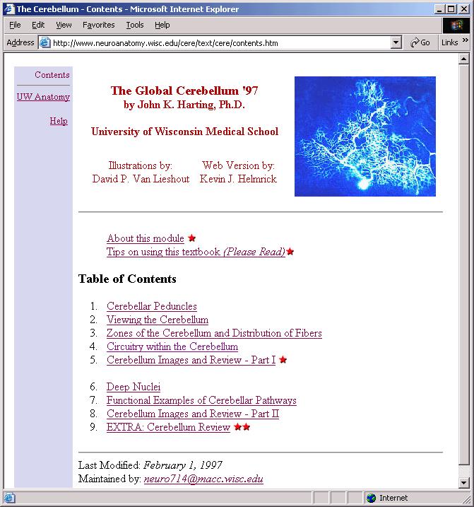
A site from the Univ of Wisconsin Anatomy Dept, with good graphics, and
sound bites!
http://www.neuroanatomy.wisc.edu/cere/text/cere/contents.htm
below is a screen shot of the index page from Univ Wisc...

Oddly, the Univ of Wisc webpage does not mention stellate cells, whose dendrites
receive input from the parallel fibers and whose axons project to and inhibit
Purkinje cell DENDRITES.
the site below has sections from human brain...
http://137.222.110.150/calnet/Cereb/page2.htm
The Society for Neuroscience background page on the cerebellum:
http://www.sfn.org/content/Publications/BrainBackgrounders/cerebellum.htm
What does the cerebellum do? The cerebellum coordinates fine movement and
is a site of motor learning, memory, and "repair". A simple point of view:
the cerebellum is responsible for coordinated movement, the basal ganglia for stationary
posture.
quotes from J.F. Stein, pp 75-80 (1985):
"The most difficult problem of all is that [the cerebellum] is an essential
node in numerous feedback pathways; hence studying the cerebellum in isolation from
other components of these loops reveals little about its true function."
"Holmes (1939) suggested a function for the cerebellum that I believe
still remains the best guess available. He proposed that the regularly repeating
units of which the cerebellum is composed may serve as 'error detectors' -comparing
the intended course of a movement with the way it actually progresses."
Where is the cerebellum? See demo: "Brain in a Jar." The cerebellum
("little brain") sits above the spinal cord, at the base of the brain,
and seems to be in its own world, with extensive sensory inputs and v. constrained
outputs. All vertebrates have some kind of cerebellum, all with the same basic
5-cell circuitry. The cerebellum of dolphins is huge, perhaps because of the
dolphin's problems with navigating in the 3-dimensional space of open ocean;
we humans have only 2-D surfaces to navigate.
Why do we care about the cerebellum? Lesions of the cerebellum affect eye
movements. The cerebellum is implicated in adaptations such as gain change of VOR
and recovery from saccadic dysmetria (Assgn 5, 9).
Cerebellar circuitry. The structure of the cerebellum is probably better
understood than any CNS region except the retina. But knowing that structure,
and knowing that the cerebellum is involved in control of movement doesn't seem
to be enough to tell us exactly what the cerebellum is doing. Compare to a region
like the retina, where it will be clear what role each cell type plays in visual
processing.
Anatomical folia diagram, from Nauta and Fiertag:
Cerebellar input:
¶ Various sensory data (visual, auditory, proprioception, joint angle,
skin pressure...) enter the cerebellum as mossy fibers mf synapsing
on granule cells. gran. The mossy fiber input can be considered "context"
because there is so much of it.
RHSC2:
p. 240 ff: vestibular information reaches the cerebellum via mossy fibers; a
few are primary fibers, but most come from the vestibular nucleus. Main projection
is to vestibulocerebellum, but can project to vermis, flocculus, and to the
DCN fastigal nuclei.
Fig. 9.27: route by which optokinetic stimulation can reach cerebellum:
retina projects to NOT (nucleus of the optic tract), then to the inferior olive
(IO). IO axons are climbing fibers.
top of page 253, and figure 9.32: passive rotation of the eye can result
in purkinje cell responses! evidence of stretch receptors in extraocular muscles?
¶ Other sensory data, somehow considered "error signals",
such as retinal slip, enter the cerebellum via the Inferior olive. IF axons "climb"
Purkinje cells bodies and act as powerful excitatory input. The climbing fibers
cf can be thought of as "teaching" or correction inputs.
Cell types
1. granule cells: The bottom layer of the cerebellum is packed with
granules, perhaps 10 billion. Each gran cell is contacted by a mossy fiber input
(1-to-1?) and each granule cell sends an axon up to the top (fiber) layer of
the cerebellum, where it bifurcates into one parallel fiber. Perpendicular
to the aptly named parallel fibers are the flat but wide dendritic trees of
2. Purkinje cells: to which the parallel fibers make excitatory contact.
About 80,000 synapses per dendritic tree. Movie of
a Purkinje cell rotating, to show the flat, wide structure. P-cells also
receive excitatory contact on their cell bodies from climbing fibers,
originating in the Inferior Olive. (The slip signal from the retina ends up
on a climbing fiber.) Purkinje cells provide the only output of the cerebellum.
Inhibiting Purkinje cells are
3. Basket cells, which send strong inhibitory signals to the P-cell
body; the basket cells, like the P-cells, receive their "context"
input from the parallel fibers. One basket cell fans out to about a dozen P-cells.
also inhibiting P-cells are
4. Stellate cells, which provide selective inhibition to parts of the
P-cell dendritic tree. Again, stellates receive their input from the parallel
fibers.
5. Golgi cells are the fifth cell type in the cerebellum. They receive
parallel fiber input (excitatory) and inhibit granule cells. One
Deep cerebellar nuclei: are the only site for P-cell output, and the
P-cells are inhibitory in the DCN. The nuclei are the fastigal, globose,
emboliform and dentate. Axons from the fastigal nucleus project to the vestibular
nuclei. Other workers find that Purkinje cells inhibit second order neurons
in the Vest Nuc and thus can affect gain of the VOR.
Connection matrix:
FROM rows, TO columns
|
|
P-cell dendrite
|
P-cell body
|
gran cell
|
basket
dendrite
|
stellate
dendrite
|
Golgi
dendrite
|
DCN
|
|
mf
|
|
|
+
|
|
|
|
|
|
pf
|
+
|
|
|
+
|
+
|
+
|
|
|
P-cell axon
|
|
|
|
|
|
|
_
|
|
basket axon
|
|
_ _
|
|
|
|
|
|
|
stellate axon
|
_
|
|
|
|
|
|
|
|
Golgi axon
|
|
|
_
|
|
|
|
|
|
cf
|
|
++
|
|
|
|
|
|
Cell numbers
Total number of Purkinje cells in human cerebellum: about 15 million.
Total granule cell count: about 10 billion. Granule cells small
(3 - 5 microns spheres) and packed together in the "bottom" layer
of the cerebellum. Try a volume calculation:
(4/3)*pi*(2 x 10-6)^3 * 10^10 = 3.35 x 10⁻⁶ m^3 = about 3 cc of volume, not unreasonable.
How can we account for the spaces between the spheres?! Actual volume probably more
like 4 cc.
At any rate, the number of granule cells is huge: perhaps half the nerve cells in
the brain are in the granule layer of the cerebellum.
If there are 10^10 granule cells and 80,000 parallel fiber synapses per Purkinje
cell, how many P cells are contacted by one parallel fiber? (each granule cell
sprouts one pf...)
ANS: there are about 1000 gran cells per P cell. 80,000/1000 ≈ 80 P cells/pf.
Effects of cerebellar lesions.
First of all, even if the entire cerebellum is removed, paralysis doesn't result.
Not so, strokes in the motor cortex, or the loss of motor function of by degeneration
of the basal ganglia (Parkinsonism); there a severe lack of movement can result.
One result: Extremity ataxia: impairs movements of the ipsilateral limbs. Test
by asking for extension of ipsi index finger, then touching finger to nose with
eyes closed: done with "marked instability" without cerebellum.
Nauta & Feirtag, p 103: "Lesions of the vermis, the midline zone of
the cerebellar cortex, and especially lesions involving the fastigal nucleus
(the most medial of the DCN) often leave the extremities unimpaired. Instead
they severely derange the movements of the trunk: They bring on trunk ataxia.
Here the patient proves unable to balance his trunk over his legs. Attempting
to walk, he staggers and tends to fall backwards."
From RHSC2: Affects of lesions:
Prolonged spontaneous nystagmus after ablation of vestibulo-cerebellum. Inability
to hold a deviation of gaze.
Cerebellar lesions can interfere greatly with smooth pursuit.
Cerebellar lesions can produce errors in the amplitude of saccades...
"...many cerebellar afflictions are remarkably similar to the effects of
alcohol poisoning."
Cerebellum and VOR gain control:
For a recent review of the role of the cerebellum in VOR gain control, see Ito
2002. In Ito's Fig 1 I think he has the sign wrong for Purkinje cell
and Golgi cell ouput: both should be inhibitory, not excitatory as drawn.
Summary
§ Role of the cerebellum in motor control
§ "Wiring diagram" of the cerebellum
§ Deep cerebellar nuclei and the vestibular nucleus
§ Effects of cerebellar lesions
