BIBS awards five Innovation Grants
The Brown Institute for Brain Science has awarded BIBS Innovation Awards to five research teams to help these groups launch new, creative research projects with great potential that are too risky and early stage for external funding sources. Thanks to a generous donation, BIBS awards up to $100,000 for one year for each project. A second year of support is also possible on a competitive basis.
In this second year of the program 19 applications were received, including two that were seeking a second year of support. Following review by a panel of researchers that included Brown faculty and experts from outside Brown, BIBS is pleased to support the following projects.
Establishing a new technique for circuit tracing and manipulation
PI: Gilad Barnea, PhD, Neuroscience
The objective of this project is to establish a new method for labeling and manipulating neural circuits – a major challenge in studying the brain. The project builds on a new technology called trans-Tango that Dr. Barnea and his team have developed and implemented in fruit flies (Drosophila melanogaster) with support from the first year of the BIBS Innovation fund. In this second year, the team at the Barnea lab will implement the technology in mice. Successful implementation of trans-Tango in mice will deepen our understanding of fundamental mechanisms that underlie brain function as well as the causes and progression of many diseases of the nervous system.
How do SLC13A5 mutations cause severe infantile epilepsy and how can we use this knowledge to develop new and novel treatments for pediatric epilepsy?
PI: Stephen Helfand, MD, CMB
This project focuses on a gene that has been linked to a rare form of infantile epilepsy. Mutations in the gene SLC13A5, which normally encodes a protein that transports citrate across cell membranes, lead to seizures in children. Dr. Helfand has studied the corresponding gene, or homologue, in the fruit fly Drosophila melanogaster and in mice in order to determine how the gene functions in metabolism and aging. He will use that background expertise to develop new animal models of seizures with the goal of identifying new treatments for these rare seizures. To start this effort in Drosophila he will replace the fly gene with the human version, introduce one of the epilepsy causing mutations, and determine whether it leads to seizures in flies. These will be the first steps that will provide proof of principle for a larger effort to identify small molecule drugs that can correct the action of the mutation and possibly shed light on the etiology and treatment of other pediatric epilepsies.
Regulation of the axonal translatome during nervous system wiring
PI: Alex Jaworski, PhD, Neuroscience
This project addresses long-standing questions about how neurons find their way to their appropriate targets as an organism develops. Dr. Jaworski will study the spinal cord of developing mice, where a select group of neurons send their cable-like axons from one side of the spinal cord to the other. Previous studies have shown that there are proteins that attract these axons toward the midline. After they cross, other proteins repel them from the midline. This project will investigate the mechanism of this axonal “switch” in response to midline signals. Jaworski hypothesizes that this switch occurs because of local changes in mRNA translation which alters protein synthesis within the axon. His team will analyze which genes are producing proteins at these two stages. The research group will then alter the timing of expression for these mRNAs experimentally and examine whether growing axons can still cross the midline.
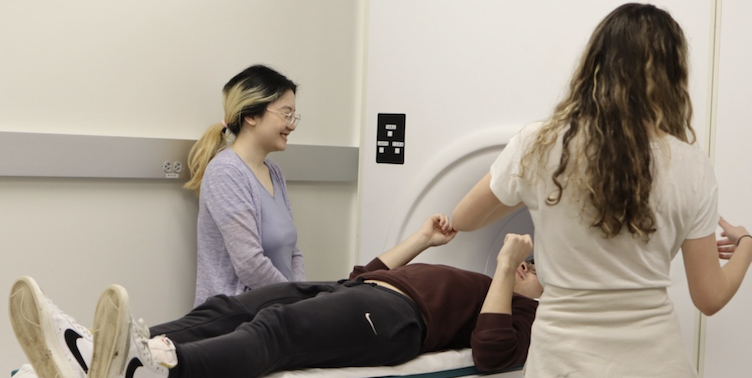
Advanced Neuroimaging of Functional Connectivity and Networks
PIs: Jerome Sanes, PhD, Neuroscience; Xi Luo, PhD, Biostatistics; Eli Upfal, PhD, Computer Science
This is a collaborative project between experts in brain imaging (Sanes), biostatistics (Luo) and computer science (Upfal) who will combine their skills to create new methods for analyzing the activity of specific neural networks in the human brain and how they change dynamically during learning. This project will develop and implement an integrated and scalable computational and statistical framework to assess activity in local and longer-range brain networks, as revealed through simultaneously recorded electroencephalography and functional magnetic resonance imaging (fMRI) signals in humans while they learn and perform action sequences. These activity maps will be integrated with anatomical information about interconnections in the brains of individual people using a specific MRI technique called diffusion spectrum imaging. The goal is to track brain activity associated with specific tasks as it flows through large regions of the brain in real time and show how it changes dynamically as people learn or change their attention.
Comparative Functional Neuroimaging for Cognitive Research at Brown
PIs: David Sheinberg, PhD, Neuroscience; David Badre, PhD, CLPS; Theresa Desrochers, PhD, CLPS/Neuroscience
This project has two important goals: 1) to establish a working protocol to carry out monkey fMRI studies at Brown; and 2) to compare brain circuits for remembering temporally ordered visual events in both monkeys and humans using fMRI. It will bring a new technology to Brown that will provide a direct means to compare functional brain circuitry in humans and non-human primates while they perform the same tasks. This is a vitally important link that is largely unavailable. There are only a few labs in the world that conduct fMRI studies with non-human primates and even fewer that attempt to use this technology to reveal functional brain pathways linked to higher cognitive processes, like remembering sequences, in both non-human primates and humans.