Myxoid liposarcoma
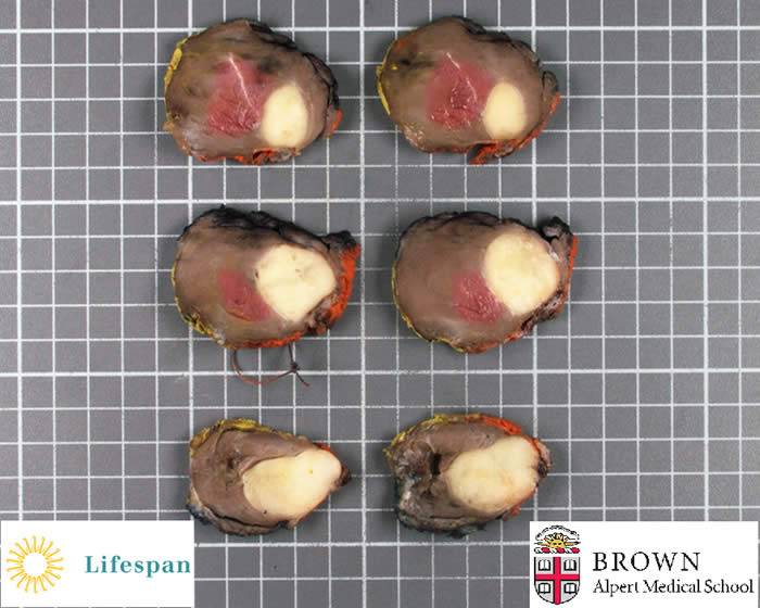
37 year old female presented with pain in the left knee and was incidentally discovered to have a mass in the anterior tibialis. The lesion was biopsied, diagnosed and she subsequently received preoperative radiation therapy followed by resection of the anterior tibialis muscle.
Cross sections reveal a yellow white, homogenous, well circumscribed mass deep within the muscle.
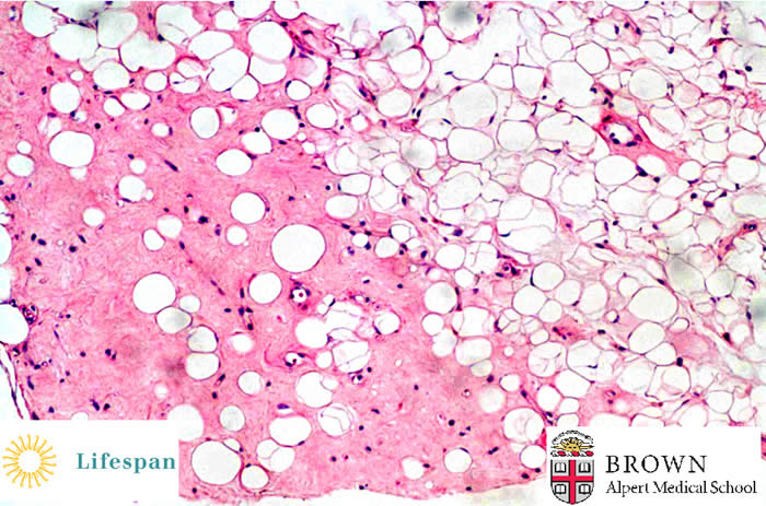
Microscopic sections reveal numerous atypical adipocytes suspended in a prominent myxoid stroma with ‘chicken wire’ capillary vasculature, characteristic of myxoid liposarcoma.
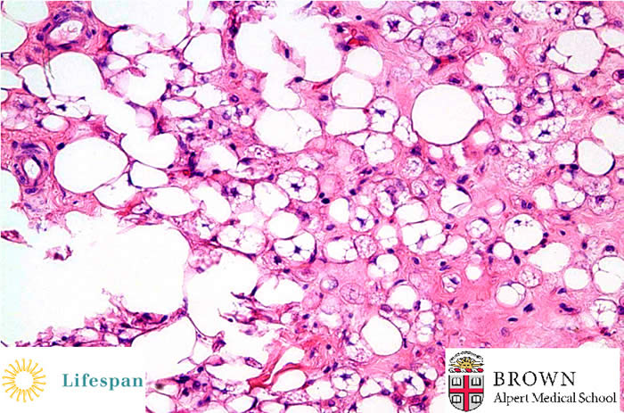
A focal area demonstrated numerous lipoblasts. No round cell component was identified in the lesion.
The hallmark cytogenetics of these two entities is t(12;16) (q13;p11) which can be found in more than 90% of all cases. Reciprocal translocation t(12;16)(q13;p11) of myxoid-round cell liposarcoma, described about twenty years ago, results in a fusion gene consisting of the 5' part of the FUS (TLS) gene and the complete coding region of the CHOP gene.
CHOP, acronym of C/EBP-Homologous protein, is also called DDIT3 which stands for DNA damage-induced transcript 3. The second translocation of myxoid liposarcoma is associated to t(12;22;20) (q13;q12;q11) chromosomal translocation, resulting in the fusion of the DDIT3 and EWS genes.
Contributed by Sonja Chen MD
Back to Bone and Soft Tissue section