Giant cell tumor of tendon sheath
Giant cell tumor of tendon sheath, also called nodular tenosynovitis, belongs to the localized variant of tenosynovial giant cell tumor. Tenosynovial giant cell tumors harbor a consistent chromosomal translocation, t(1;2)(p13;q37). The resultant fusion gene has a promoter of collagen type VI alpha-3 gene (Col6A3) and a coding sequence of colony-stimulating factor 1 (CSF1). Col6A3 gene is constitutively expressed in the synovial lining of joints, tendon sheaths, and bursae. Driven by active promoter, the fusion gene produces a large amount of CSF1 which is a potent chemoattractant for macrophages. As a result, numerous histocytes infiltrate the tumor with evenly dispersed multinucleated giant cells as well as lymphocytes and other inflammatory cells.
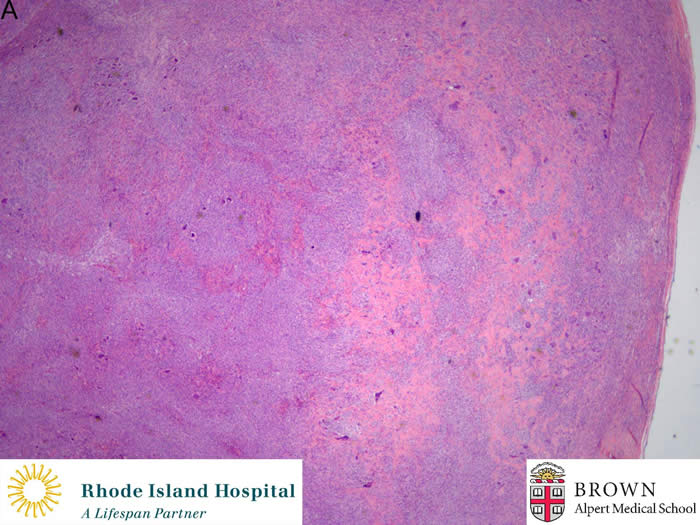
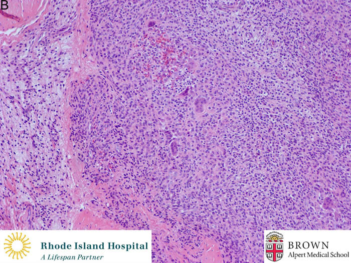
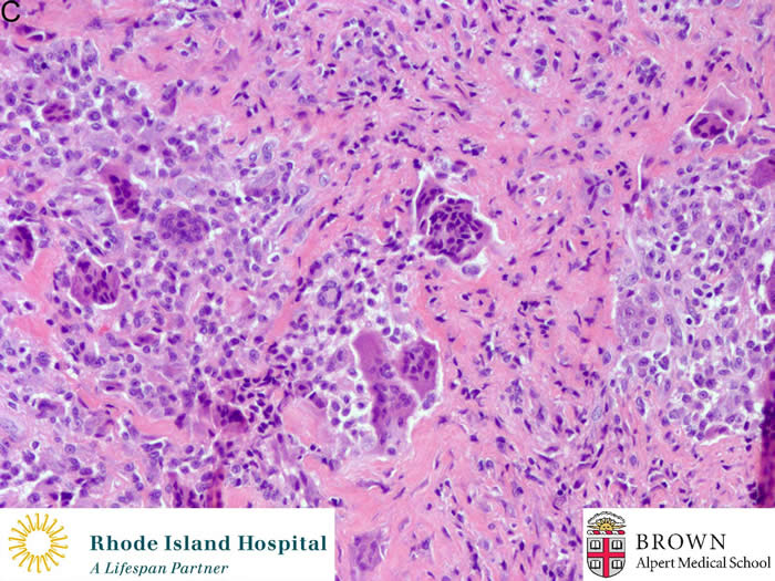
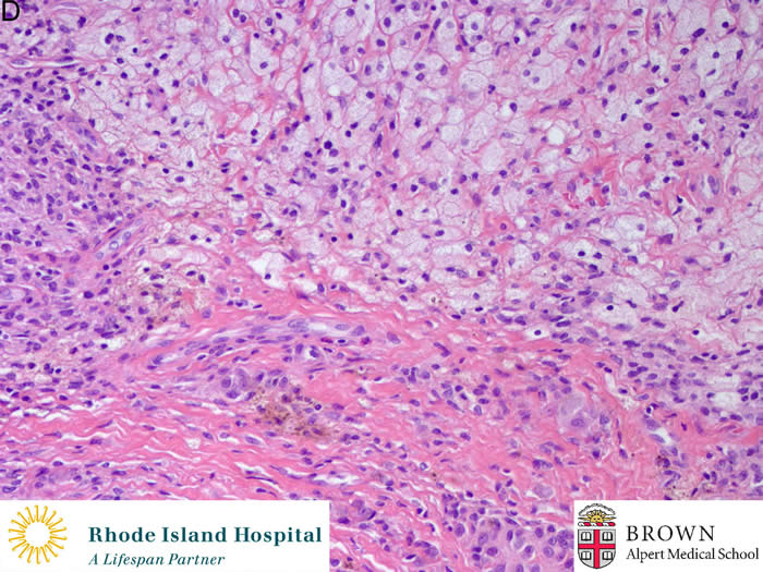
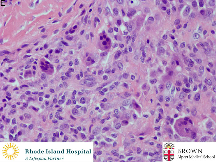
Giant cell tumor of tendon sheath is usually a small, circumscribed, lobulated mass (Figure A). The lesional cells only account for 2%-16% of the cells in the mass. They are polyhedral, moderately sized, modified synoviocytes. Foamy macrophages and hemosiderins are frequently seen (Figure D). The lesion has cleft-like spaces (Figure C&E) and a scattered mitotic figure may be found.
Although giant cell tumor of tendon sheath is a benign lesion, local recurrence is about 20% after surgery.
Contribute by Michael Chaump M.D.