Osteogenesis imperfecta congenita (type II)
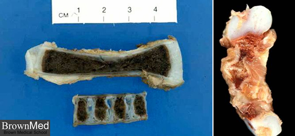
The photo on the left shows longitudinal sections of a femur and the spine from a normal mid-gestation fetus.
On the left is a femur from a patient with OI type II. It is short and curved and is the site of multiple fractures.
1 minute clinical correlation