Tetralogy of Fallot (2)
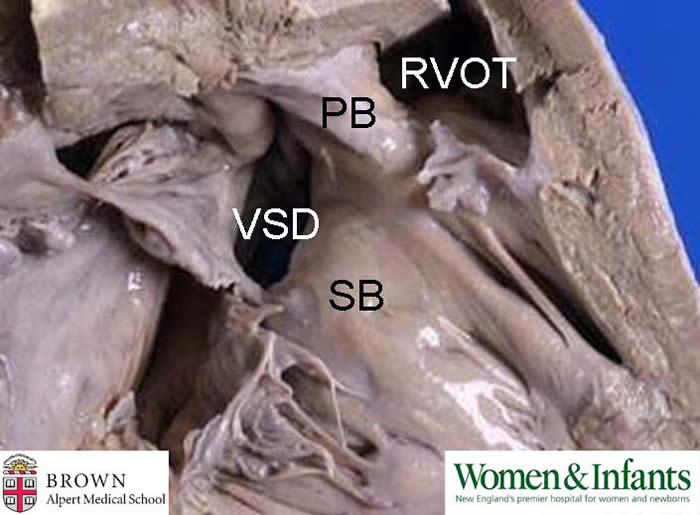
This is a photo of the right ventricular outflow tract in an adult with tetralogy of Fallot (ToF). The anatomical basis of ToF is an anterior malalignment of the parietal band (PB). As a result of this malalignment a conoventricular septal defect (VSD) is created and the right ventricular outflow tract (RVOT) is narrowed. The pulmonary valve may or may not also be stenotic or even atretic.
Clinical correlation: In addition to a VSD and narrowing of the RVOT the other features of tetralogy of Fallot (ToF) are right ventricular hypertrophy and overriding of the aorta. In the fetus, of course, “right ventricular hypertrophy” is the norm. The normal anatomy of the heart includes over-riding of the aorta. Overriding is exaggerated in ToF and in another anomaly closely resembling ToF, double outlet right ventricle (DORV).
ToF is often now discovered in utero. A newborn with ToF will quickly become cyanotic after birth when the ductus arteriosus closes. The amount of obstruction in the RVOT or pulmonary valve strongly correlates with the severity of symptoms.
Contributed by Dr. Calvin Oyer