Tetralogy of Fallot (3)
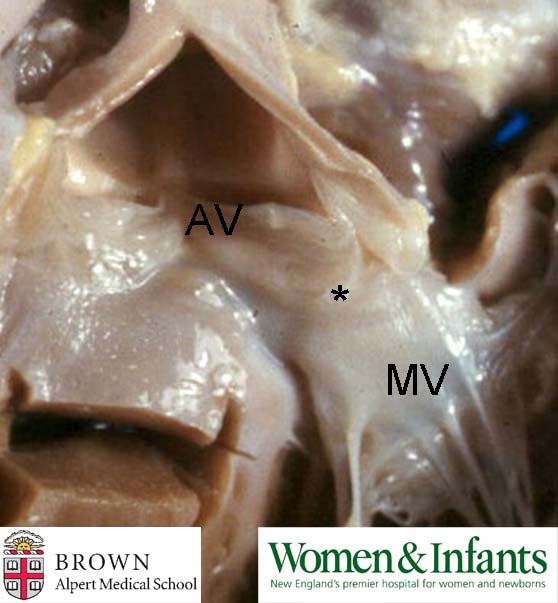
This is a photo of a normal left ventricular outflow tract (LVOT) showing fibrous continuity between the anterior leaflet of the mitral valve (MV) and the noncoronary cusp of the aortic valve (AV). An anatomical distinction can be made between tetralogy of Fallot (ToF) and double outlet right ventricle (DORV) by examination of the left ventricular outflow tract (LVOT). In ToF there will be normal fibrous continuity between the aortic and mitral valves as shown in this photo. In DORV such continuity will be lacking due to interposed muscle; i.e. in DORV there are bilateral conuses whereas in ToF there is a conus only on the right.
Contributed by Dr. Calvin Oyer