Glioblastoma multiforme (GBM)
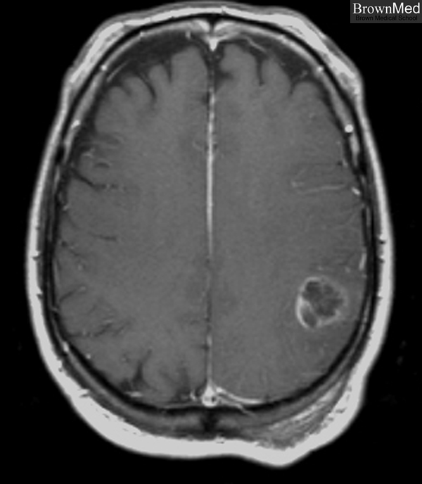
Axial view of T1-weighted MR image of the brain prior to surgical resection demonstrating
a 2.2 x 2.3 x 2.3 cm heterogeneous enhancing mass in the left inferior parietal lobe.
1 minute clinical correlation
Previous | Home | Next
Back to neuropathology section