‘ScanFest’ sessions teach students the ins and outs of using an MRI machine for research

As part of a class taught by Brown neuroscientist David Badre, undergraduates embrace the rare opportunity to conduct experiments and engage in research with state-of-the-art MRI technology.

PROVIDENCE, R.I. [Brown University] — On a recent day in the MRI Research Facility in the Sidney Frank Hall for the Life Sciences on Brown’s campus, it wasn’t too difficult to tell which undergraduates were planning to go inside the magnetic resonance imaging (MRI) machine.

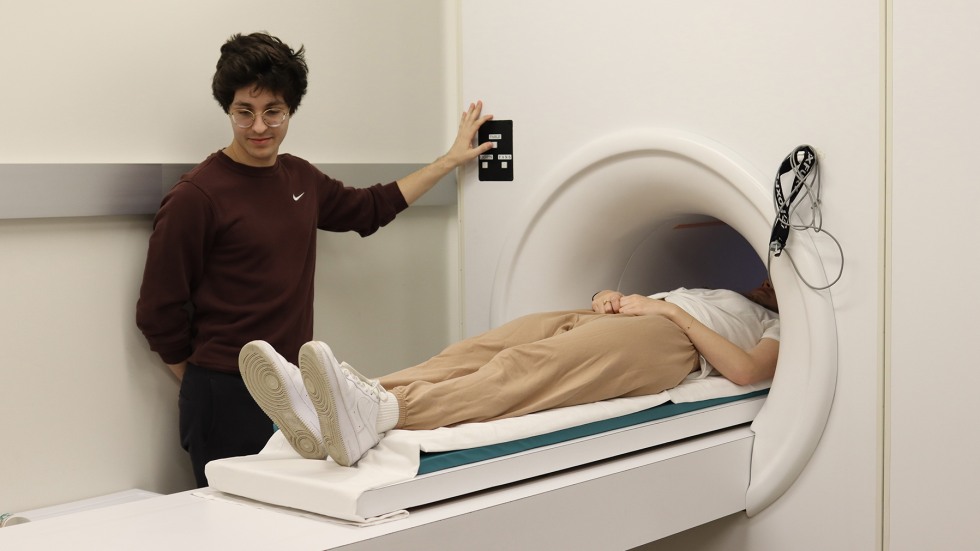
While most of the assembled students were dressed in sweatpants and T-shirts, the ones who volunteered to go into the machine to have their brains scanned were completely metal-free: No jewelry, no belts, no shoes. They were scrupulous about this, since screening MRI participants and knowing what one can and can’t wear inside the powerfully magnetic machine is part of the course they’re taking.

The eight students gathered at the research facility are enrolled in the Functional Neuroimaging course taught by David Badre, a professor of cognitive, linguistic and psychological sciences who is affiliated with Brown’s Carney Institute for Brain Science. The goal of the spring course is to train students in the practice and use of MRI to advance cognitive neuroscience research and it provides students with the opportunity to go inside the MRI machine, operate it and participate in other key ways. 

“We are scientists, we work in the lab, and this MRI facility is our lab,” Badre said. “So we need specific training on how to properly use this equipment.”

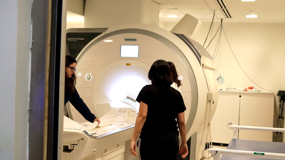
For two Saturdays in April, the class convened inside the facility, a 3,000-square-foot research suite that features a state-of-the-art Siemens 3 Tesla PRISMA scanner. While the facility is a resource available to researchers at Brown and Brown-affiliated hospitals, it’s not commonly used by undergraduates.

The two interactive class sessions, which have come to be known as ScanFest, are a chance for students learn how to prepare research subjects, set up the machinery and monitor activity in the MRI control room.

“It’s very rare for a university to donate time to undergraduate students for scanning practice and training,” Badre said.

The scanning sessions have always been a high point of the course, and they were even more highly anticipated this year:  This was the first time that ScanFest has been held since 2020, when Badre adopted a remote format for the class in order to comply with University safety guidelines during the COVID-19 pandemic.

“By coming in here and using fMRI, which is functional magnetic resonance imaging, we teach the practical science in a very serious and hands-on way, but it also lets students be curious and experience the joy of scientific discovery,” Badre said.

Getting the most out of the MRI machine

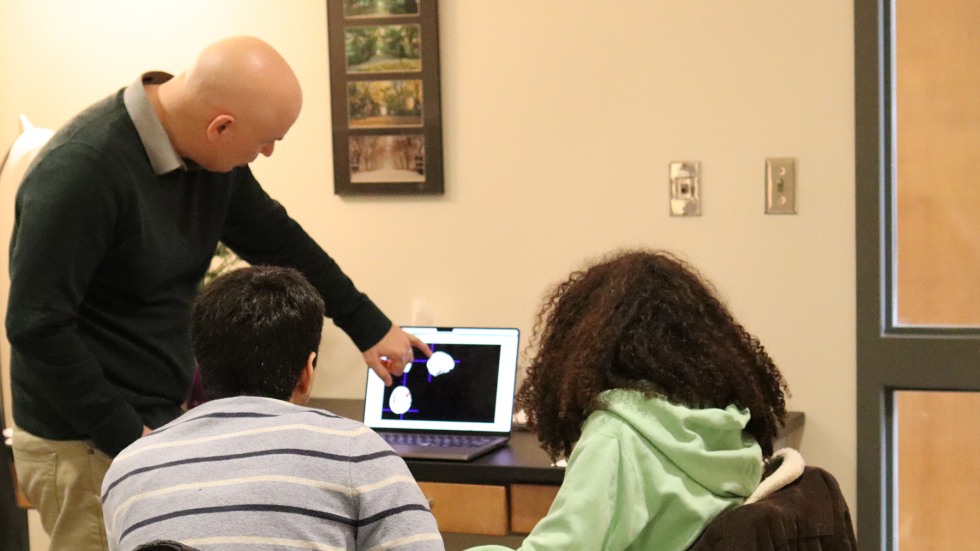
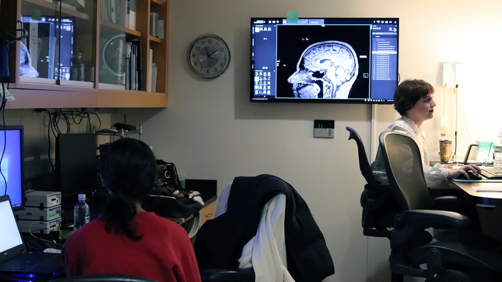
As part of the course, the students design an experiment that uses functional magnetic resonance imaging — fMRI measures brain activity by detecting changes associated with blood flow. Across the two sessions, eight or so students volunteer to be research subjects and go inside the machine to perform cognitive tasks while the having their bran activity recorded. Lynn Fanella, the MRI technician who manages the Brown facility, attends both sessions to offer students a hands-on lesson in operating the machine and reading its output.

The course also teaches students how to design a successful cognitive research experiment. fMRI is an exciting method that allows researchers to visualize the brain in a non-invasive way, Badre said, but it’s still just a tool, and getting answers depends what questions are asked and how they’re presented.

The experiments need to be relatively simple so that the students can get clear answers from the scans, said Badre, who has been teaching the class since 2008. 

“Within the restrictions we give them, the students come up with very creative ideas,” Badre said. “The thing about these scanning sessions is that when they run these experiments, they’re going get some sort of outcome in the neuroscience data — and that’s what’s exciting.”

Students explore responses to disgust, AI-generated images and more

The groups in this year’s course came up with a range of distinct experiments, including whether the brain processes photographs of faces in the same way that it processes AI-generated images of faces; how the brain reacts to an image of a farm animal versus an image of a human; an exploration of  visual and perceptual activity when a person views a familiar location and when they imagine a familiar location; and what happens inside the brain when a person views something that evokes disgust.

Preparing for ScanFest started well before the students arrived at the MRI facility.

The groups needed to look for appropriate images to show their research subjects. For example, there are different ways to provoke feelings of disgust, and through researching images, students realized that their images needed to be consistent and free of distracting elements. They decided to show plates of pleasantly appetizing food as well as unappetizing images, such as a piece of rotting fruit covered by ants.

Brown senior Hamzah Chamdia’s group was exploring how the brain processes images of sheep and other farm animals differently than those of humans. Chamdia, who is concentrating in cognitive neuroscience, noted the challenges of preparing 250 images for the experiment: once his group found images of farm animals, they spent many hours standardizing not only the sizes of the images but also the layouts (for example, they edited out barns, fences and fields so that all images of animals and people were presented against the same white background).

Students submitted research proposals, solicited feedback from classmates and met with Badre to prepare. Class assignments were structured to help students understand what they’d be looking at in terms of brain structure and activity. After collecting the data, the students perform statistical analysis and inference. At the conclusion of the course, they’ll present their findings to the rest of the class.

Chamdia appreciated learn how to advance research with the fMRI machine.

“It’s a really great opportunity because not every university has this for undergraduates,” he said.

Unlike many of the students in the course, Brown senior Elizabeth Doss has also worked in the MRI suite in her role as a research assistant for cognitive neuroscience research projects in Badre’s lab. Doss has helped scan participants to collect data and has analyzed that data, as she’ll be doing with her group — the students analyzing disgust — as part of the Functional Neuroimaging course. Doss said she enjoyed the comprehensive approach to learning about the MRI and what it can do.

“My mentor recommended this class because I was trying to learn a lot about how MRI works on my own,” Doss said. “I’d done a lot of research of MRI physics and how to best process data, and David Badre is an expert in that field. I wanted to take this class to fully understand what I was working with.”

The course provided Doss and her group with something unique: the freedom and control to design and run their own experiment. From coding tasks to running the scanner to pre-processing images and analyzing data, the students are owning every aspect of their projects.

“It’s just a different kind of process than assisting with an ongoing study designed by a Ph.D. student or faculty researcher,” Doss said. “We do everything in this class. I’m glad I can be a resource for other students, but I’m still constantly learning. There’s so much that I’ve been able to take out of the class.”

Doss had heard about the course during her sophomore year and eagerly awaited her chance to enroll.

“I was really happy to be able to take this class in my senior year, before graduating,” she said, “And it’s been a great experience.”