Peripheral Blood Counts |
|
RBC |
2.72x10^12/L |
Hemoglobin |
10.1 g/dL |
Hematocrit |
29% |
MCH |
37 pg |
MCV |
106.3 fL |
MCHC |
34.8 g/dL |
WBC |
3.0 x 10^9/L |
Absolute neutrophil count |
0.5 x 10^9/L |
Absolute lymphocyte count |
2.4 x 10^9/L |
Aboslute moncyte count |
0.1 x 10^9/L |
Absolute eosinophil count |
0.1 x 10^9/L |
Absolute basophil count |
0 x 10^9/L |
Platelets |
44 x 10^9/L |
MPV |
8.3 fL |
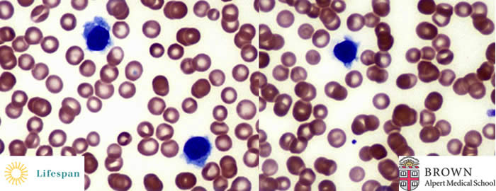
Peripheral Blood shows a subset of atypical lymphocytes with hairy-like cytoplasmic projection.
Atypical lymphoid cells are positive for TRAP
Bone Marrow Aspirate |
|
Blasts |
0.10% |
Promyelocvtes |
0% |
MetaÍmyelocytes. bands, segmented neutrophils |
2.20% |
Erythroid precursors: |
13% |
Lymphoid cells: |
82.20% |
Monocytes: |
0.80% |
Plasma cells: |
1.70% |
Eosinophils: |
0.30% |
Basophils: |
0.10% |
Mast cells: |
0.30% |
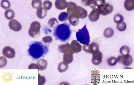
Bone marrow aspirate is remarkable for 82.2% lymphoid cells many with moderate to large amount of cytoplasm and irregular cytoplasmic hairy-like projections in a large subset.
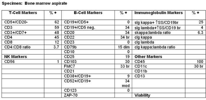
Flow Cytometry shows kappa light chain restricted B-cell population co-expressing CD20, CD103, CD11c, CD25.
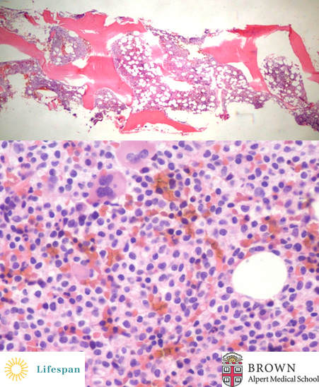
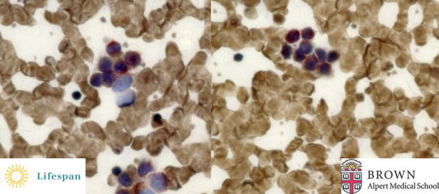
Bone Marrow Biopsy is hypercellular for patient's age (70-80 %) and remarkable for extensive replacement of the bone marrow space by a population of neoplastic lymphoid cells which comprise approximately 80-90% of the nucleated bone marrow cells.
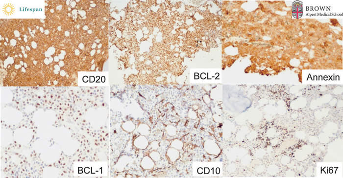
There is a large CD20+ ( >85% of the nucleated bone marrow cells), Annexin A1+, BCL2+, variably Cyclin D1+, CD10 negative neoplastic B-lymphoid population with a rather low proliferation rate (less than 5%) in the bone marrow.
The diagnosis of this case is: Hairy cell leukemia with extensive bone marrrow involvement.
Hairy cell leukemia is a rare neoplasma of small mature B lymphoid cells (post-germinal center stage, arrested at some point during isotype switching), comprising 2% of lymphoid leukemias. Patients are predominantly middle-aged to elderly adults with a median age of 50 years. Tumor cells are located predominantly in the bone marrow and spleen with a small number of circulating "hairy" cells.
The bone marrow effacement is of variable extent. The primary pattern is interstitial or patchy infiltration of widely-spaced lymphoid cells with oval or indentaed nuclei with abundant cytoplasm and prominent cell borders taking a "fried-egg" appearance. An increased reticulin fiber is common and often results in a "dry-tap".
Hairy cell leukemia is uniquely sensitive to either alpha-interferon or nucleosides such as pentostatin and cladribine. Anti-CD20, anti-CD22, and anti-CD25 are the new agents available recently of variant indications. Prolonged remission may also result from splenectomy, but this is uncommon. The overal 10-year survival rate exceeds 90%.
Contributed by Jianhong Li, MD and Dr. Diana Treaba
Back to Hematopathology Section