Aspergillosis
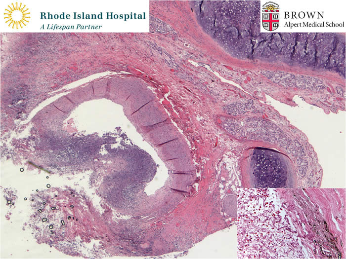
This pulmonary artery section is from a 51 year old man with a history of asthma and a 15 year history of known sarcoidosis. He had been on steroids for a long time and was recently receiving antibiotics. He died suddenly during an episode of severe hemoptysis. This microphoto shows necrosis of the pulmonary artery wall with rupture. The inset shows the presence of invasive aspergillosis.
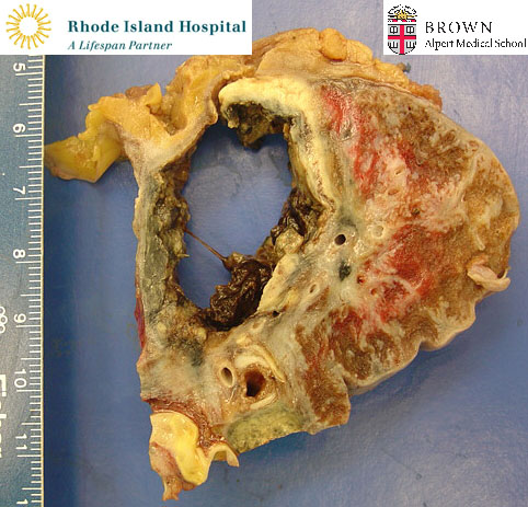
This inserted gross photo, from the same patient as
the previous slide, is of the right lung near the hilum. It shows a large
cavitary lesion. Microscopically, numerous aspergillus hyphae were noted in
necrotic material lining the wall of the cavity.
Aspergillosis can be separated into colonizing and
invasive forms. The colonizing form is represented by the formation of
aspergillomas in which the fungus grows in pre-existing cavitary lesions.
Invasive aspergillosis represents an opportunistic infection which occurs in
immunocompromised patients. It causes necrosis of tissue and tends to invade
blood vessels. In this case it resulted in fatal hemoptysis.
Contributed by Ronald DeLellis, MD and Wei Tian, MD