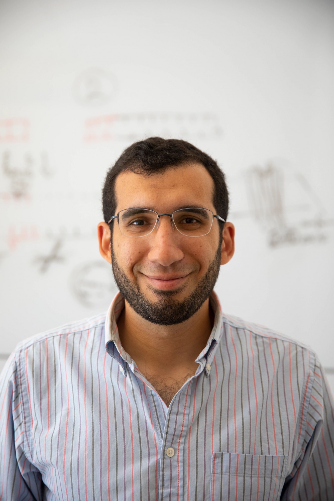
PROVIDENCE, R.I. [Brown University] — For all of its mind-blowing capabilities, the human brain remains somewhat of an enigma. Even with the most advanced imaging tools, it’s still difficult to clearly and easily identify brain processes as they’re happening. But tracking brain activity in real time is crucial to understanding brain function, said Ahmed Abdelfattah, an assistant professor of neuroscience at Brown University.
With a new five-year, $1.5 million New Innovator Award from the National Institutes of Health’s High-Risk, High-Reward Research program, Abdelfattah will work to develop molecular tools to allow researchers to “see” the brain in action. His project aims to illuminate brain communication through developing new classes of genetically encoded tools, including fluorescent reporters and tracers, that allow for mapping and manipulating brain circuits.
“We're translating biochemical signals in the brain into light so that we can use a microscope to visualize what’s happening in the brain of living animals while they’re performing certain behaviors,” said Abdelfattah, who is affiliated with Brown’s Carney Institute for Brain Science.

Brain circuits are dynamic networks of neurons that process information in the form of electrical and chemical signals to form memories and shape behaviors. Abdelfattah will repurpose naturally occurring proteins to show those signals as they’re happening in the brain. He will genetically engineer cells to express protein probes that will fluoresce, or light up, during certain activities or when detecting specific neurochemicals. These protein probes change color and brightness when the neurons fire. The cell will express the probe as it expresses its own proteins, allowing for a relatively noninvasive method of recording the precise timing of actions that have been previously undetectable.
The genetic, or “optogenetic,” feature of the probes is key, Abdelfattah said. Not only does it improve accuracy and avoid the need to inject probes via invasive or toxic methods, but it allows for precision control.
“This will give us the genetic ability to target the specific cells in which we’re interested,” Abdelfattah said. “That’s extraordinarily useful. For example, if we only want to label, say, the inhibitory inter-neurons of the second layer of the cortex of a mouse, we can do that.”
Current imaging tools only capture what the brain is doing at a particular time, Abdelfattah said, comparing them to a camera with a normal shutter speed. “You can take an image of what happened in one second, but during that time the brain could have made hundreds of unseen and unrecorded computations,” he explained.
In addition, the cellular resolution of the image may not be very high. Abdelfattah is working to create tools that can capture one frame every millisecond, or 1/1,000th of a second, with vastly improved clarity of cellular images. “High spatial and temporal resolution is how I would summarize it,” he said.
The fluorescence generated by the optogenetic tools can be easily measured, Abdelfattah said, allowing scientists to unravel the functional basis and causes of neuronal disorders at a level of detail that has not been accessible to date. In the future, this could be used to develop new treatments for such disorders. For example, one of the molecules for which he is trying to make probes is a naturally occurring opioid neuropeptide. The resulting images will expand understanding of how these types of chemicals affect circuits at the cellular level and hopefully inform the eventual development of addiction treatments.
Abdelfattah’s proposal notes many other examples of types of probes, as well. “We want to understand how brain circuitry works and hope that this will be useful for treatment of patients in a more precise manner in the future,” he said.
While Abdelfattah just joined the Brown community earlier this year and is still in the process of setting up his lab, he said that he’s already been able to test some of his molecular tools in the labs of other neuroscientists at the Carney Institute.
“It’s exciting to be part of the neuroscience department at Brown, a unique and fun place to talk with like-minded people who are thinking about big problems in brain science,” he said.
Abdelfattah is one of 64 “exceptionally creative scientists” from across the nation to receive a New Innovator Award, which supports unusually innovative research from early career investigators.
Each year the NIH recognizes trailblazing new research through four awards in the High-Risk, High-Reward research program. The program catalyzes scientific discovery by supporting highly innovative research proposals that, due to their inherent risk, may struggle in the traditional peer review process despite their transformative potential. Funding for the awards comes from the NIH Common Fund, NIGMS, National Institute of Mental Health and the National Institute of Neurological Disorders and Stroke.