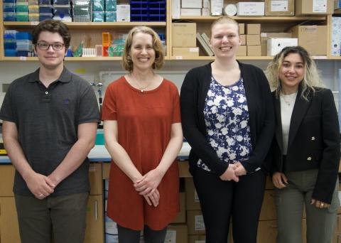
Students knock it out of the park thanks to one Carney lab’s ethos of encouragement
Diane Hoffman-Kim’s lab publishes on stroke therapy screening, showing that their “mini-brains” can serve important purposes in disease research.
Rachel McLaughlin, a fifth-year Ph.D. student in the Hoffman-Kim lab, was looking at her phone, seeking some distraction, when she saw a new email in her inbox. It was from the journal In vitro models in response to a paper she had sent them three months ago. The paper was the fruit of years of labor on McLaughlin’s part and a large component of her dissertation.
In the paper, McLaughlin described how to “test potential stroke treatments in a dish” using cortical spheroids. A flagship product of the Hoffman-Kim lab, cortical spheroids are three-dimensional spheres of rodent brain cells no bigger than the tip of a mechanical pencil. Casually referred to within the lab as “mini-brains,” cortical spheroids model actual brains in that they have the same cell types, neural communication, tissue stiffness and cell density. But what makes them different from true brains is an added boon to researchers: their tiny size and how rapidly they mature means the lab can make, maintain and conduct experiments on thousands of mini-brains at a time, quickly and cost-effectively.
McLaughlin had devised a method for depriving the mini-brains of oxygen and glucose in order to induce a stroke-like injury. She had implemented a way to analyze them — a much more difficult accomplishment in three dimensions than analysis of flat cells — and established that the cells in the injured mini-brains were struggling to communicate with each other and showed signs of inflammation just as cells in a stroke-injured brain would. Finally, she had tried pre-treating the mini-brains with a drug called N-acetylcysteine, which has shown promise in clinical trials, and discovered that the levels of overall cell metabolism and health were higher in pre-treated mini-brains despite experiencing a stroke-like injury.
Not only had McLaughlin used the mini-brains to make an exciting leap forward in treatment possibilities for stroke, she’d accomplished something her lab hadn’t yet achieved: showing that these spheroids, so far notable for their novel modeling, could serve important purposes in disease research.
McLaughlin was delighted to read that In vitro models had responded positively to the paper. However, the journal’s peer reviewers wanted more.
To determine the pre-treated mini-brains’ overall health, McLaughlin had measured ATP, or adenosine triphosphate, an organic compound the body uses for energy. While ATP is a good indicator of a mini-brain’s health across the board, it is not as specific as calcium imaging, which gives a scientist a more detailed picture of what is happening in individual cells. Could McLaughlin run her experiments again, the reviewers asked, this time using calcium imaging? Could she do this across the multiple outputs she used to characterize the stroke-like injury?
McLaughlin could not. Not anytime soon. She was looking at her phone from a hospital bed, where she was busy with labor of a different sort, giving birth to her daughter.
***
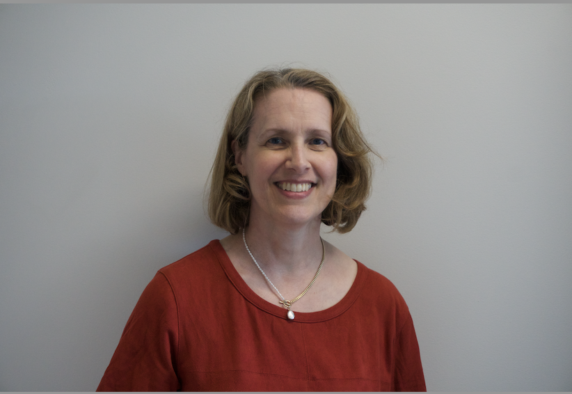
When Diane Hoffman-Kim saw the revision request land in her inbox, followed almost immediately by a picture of McLaughlin’s newborn daughter, she sprang into action.
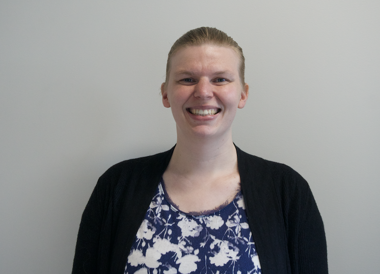
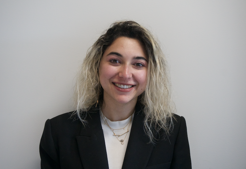
Together with Hoffman-Kim, McLaughlin had trained and nurtured many members of the lab, including Ilayda Top and Harrison Katz. Both were poised at the start of a very important academic year: the beginning of an accelerated one-year master’s program in biotechnology for Top, and the final year of an undergraduate degree for Katz. Each was preparing to begin their thesis projects. But the lab didn’t need these projects as urgently as it needed mini-McLaughlins.
Hoffman-Kim asked the two if they would be comfortable changing the focus of their theses.
In this unexpected request, Top and Katz saw an opportunity. Neither had participated in a paper revision process before. Rather than working under McLaughlin’s supervision, they would be carrying out the experiments and coding with much more independence.
The pair agreed to Hoffman-Kim’s proposition with enthusiasm.
There was a deadline to hit.
***
Using a pipette, Top set the tiniest amount of fluorescent dye atop each of the 96 mini-brains nested in a customized plastic culture dish about the length and width of her hand. She had done her undergraduate degree at Brown, in the Hoffman-Kim lab, and was immediately drawn to stroke research due to her own personal connection to the disease. But nobody had taught her how to do calcium imaging before.
Anytime a neuron communicates with another neuron, it creates an influx of calcium. This is what makes calcium imaging an excellent way to show communication—or the lack thereof—between cells. By introducing a dye that becomes bright green when it binds to calcium, a scientist can show whether the communication between cells is healthy (lots of calcium influx and therefore lots of green signal) or unhealthy (less calcium influx and less green signal). However, the process can be exceptionally difficult to perform in live cells because of how sensitive living mini-brain cells are to changes in the environment.
Hoffman-Kim was protecting McLaughlin’s time while she was away on leave, fielding most questions that came up and acting as the sole point of contact between McLaughlin and the lab. To teach Top how to do the calcium imaging however, McLaughlin opted to train Top herself over the course of a single two-hour Zoom call.
Soon, Top was also contending with another challenge. Gas contamination among U.S. suppliers had created a CO₂ shortage. CO₂ was an essential ingredient for growing and maintaining mini-brains. Critical to the lab’s incubators, it provided the mini-brains with a stable environment. The lab’s large incubator, which required massive amounts of CO₂, was not going to be an option. Any incubation the lab needed to do was now only possible in a tiny incubator the size of a hotel-room fridge, located down a hall and in a separate room from the sterile area where lab members worked with live cells.
McLaughlin had once told Top that working with the mini-brains was like caring for very needy children. Because they were growing in a non-natural environment, they required far more nourishment than ordinary brain cells ever would. But tending to the mini-brains under the circumstances Top now found herself in was even more of a delicate calculus. The cells needed to be kept at body temperature (37°C) to develop properly and to stay alive throughout experiments. The amount of CO₂ necessary fluctuated depending on how many mini-brains were in the incubator and how often the incubator door was opened. Mini-brains could be damaged if they were jostled as more trays were loaded or removed, or on their long journey from one workspace to another.
The lab decided Top should be the only person on the team allowed to use the incubator.
Top finished staining the 96 mini-brains. She took them out from under the sterile hood, maneuvered around her labmates in a space no bigger than a walk-in closet, opened the door, walked down a long hallway, opened another door, walked through the main Hoffman-Kim lab space, and—very, very carefully—opened the incubator.
***
Katz opened a video file on the lab’s computer. The file showed a spheroid which Top had grown, induced a stroke-like injury in, and exposed to dye which turned green when bonded with calcium. He compared the green fluorescence in regions of interest with the file’s baseline background level of fluorescence. Then, with his cursor, he began manually circling every cell or area of interest, marking it for further analysis.
In addition to the McLaughlin-shaped hole in the calcium imaging process, there was a similar missing piece in terms of its analysis. Luckily for the lab, Katz had spent his entire undergraduate career with this work. He joined not as an upperclassmen as most undergraduates do, but very nearly on his first day of Brown, when chance placed him in a first-year orientation group led by Hoffman-Kim and he talked his way into a seat at her lab meetings. From there, he began working with Jess Sevetson, a former Ph.D. student who created the coding that enabled the lab’s software to analyze calcium imaging on a frame-by-frame and pixel-by-pixel basis.
No matter how good someone is at calcium imaging analysis, however, they have to reckon with the fact that it takes a very long time to do precise work. Looking at 60 or 70 cells in each spheroid, and manually circling hundreds of cells across dozens of spheroids typically took Katz between 20 to 30 hours.
A journal can refuse to publish a paper at any point before publication. Hoffman-Kim had written to In vitro models once, detailing the experiments needed to answer the reviewers. She’d written again, explaining about the CO₂ shortage. The lab had already received two deadline extensions, first to January 25, 2023, and then to February 25. Asking for a third was unthinkable.
20 to 30 working hours later, Katz finished circling areas of green fluorescence and powered up a second software program. The program was called BigDataConcat, and it was notorious throughout the lab for being difficult to work with because of—as the name emphasized—how much data it generated. However, BigDataConcat was also the best way to get the reviewers detailed answers to the questions they’d asked McLaughlin about communication between cells in the pre-treated mini-brains. Questions like, if one cell fires, do other nearby cells also fire? How many of the cells are firing? How frequently do the cells fire?
Katz began coding BigDataConcat to obtain these answers and to turn them into graphs that would become figures in the revised paper. He didn’t have much time left; his deadline was looming even sooner than the group’s. He needed to get all the outputs to McLaughlin, who was coming back from her leave on January 16. She was the only person who could vet the data and verify its accuracy.
***
For the first time in months, McLaughlin took a seat at her computer to begin examining the data. Her daughter, Amelia, was now four months old. McLaughlin was about to find out if the revisions had gone according to plan.
Throughout her leave, McLaughlin had never felt any external pressure to be involved. At the same time, this research was deeply important to her. Working with Hoffman-Kim, the two scientists came up with a system that let McLaughlin focus on her family while staying in the loop with the lab. She answered questions on her own time frame, which was sometimes at 2:00 a.m. while Amelia slept, or during parts of the day when her husband, Justin, was on baby duty.
McLaughlin started opening files. She was performing quality control, checking if a cell had moved during Top’s imaging process, if the data had proved too unwieldy for Katz’s coding. The final, final, final revision deadline of February 25 was just over a month away, too soon to perform another experiment. But McLaughlin wasn’t worried.
She wasn’t worried because of the ethos of the Hoffman-Kim lab, which she’d noticed from the very beginning of her time as a Ph.D. student. At lab meetings, everyone — from the newest undergrad to the most senior grad student — were encouraged to speak, ask questions, and receive and provide feedback. Its leaders, Hoffman-Kim and lab manager Liane Livi, made the decision to work together in 2001, while their two and four year-olds played together under the table of a Wayland Square cafe. As mothers of young children, the pair intentionally built a workplace that allowed flexibility, and where voices were elevated without reservation.
McLaughlin 100% trusted Top and Katz had done the calcium imaging work exactly the way she would have done it.
She was right.
***
“Cortical Spheroid Model for Studying the Effects of Ischemic Brain Injury” was published in In vitro models on March 29, 2023. The portion of the paper dedicated to the pre-treatment of stroke-injured mini-brains with N-acetylcysteine robustly established that pre-treatment not only preserved overall cell health, it preserved communication between cells.
For her work on the project, Top was awarded the Outstanding Academic Accomplishment by a Master's Student award, which was presented to her at her commencement in front of all graduating master’s students.
For his work on the project, Katz received the Richard J. Goss Prize in Experimental Biology, a prize across all graduating seniors in the life sciences which was presented at commencement in front of the entire class of 2023.
McLaughlin was selected to participate in Research Matters 2023, a widely-publicized evening where just twelve students annually from across the entire Brown graduate student community present their research in the style of a TED Talk, and make a case for why it matters.
She knocked it out of the park.
*Acknowledgments are due to Amanda Laguna and Christien Hernandez, who, over the pandemic shut-down, developed analysis protocols necessary for the subsequent work described in this article.
Rachel McLaughlin presenting on "Testing stroke therapies in a dish" for Research Matters.


